Alternate header for print version


Licensing
Attribution Non-Commercial; No Derivatives:This image is licensed under a Creative Commons Attribution, Non-Commercial, No Derivatives License. View License Deed | View Legal Code
tweet  
*CIL – Cell Image Library accession number. Please use this to reference an image.

CIL:13091*  Cite 
Description

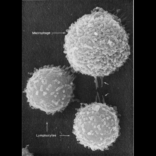
This image shows two different types of white blood cells from a mouse which are essential for mammalian immune response to protect from infection. The larger cell is a macrophage and the smaller two are leukocytes such as t-cells. Here, the cell types differ in surface texture (or morphology); the macrophage has surface folds and dense microvilli, whereas the surface of the lymphocytes is comparatively smooth with sparse microvilli. Both cell types respond to neighboring cells by extending longer microvilli (arrows). The folds and microvilli protrude due to f-actin networks under the cell membrane. Image courtesy of Emma Shelton and Jan Orenstein appears as Figure 34 from Chapter 2 (Specializations Of The Free Surface) of 'The Cell, 2nd Ed.' by Don W. Fawcett M.D. A PDF copy of the accompanying chapter is available on the ASCB's BioEDUCATE website.

Technical Details

Scanning electron micrograph.

Biological Sources
NCBI Organism Classification
Mus musculus
Cell Type
macrophage
lymphocyte
Cellular Component
microvillus
plasma membrane
Biological Context
Biological Process
immune system process
cell projection organization
Attribution
Names
Emma Shelton
Jan Orenstein
Published
ISBN 0721635849
Link
Fawcett's "The Cell, 2nd Ed."
Citation
Digital Object Identifier (DOI)
doi:10.7295/W9CIL13091
Archival Resource Key (ARK)
ark:/b7295/w9cil13091
Grouping This image is part of a group.
Imaging
Image Type
scanning electron microscopy (SEM)
Image Mode
illumination by electrons
secondary_electron imaging
Parameters Imaged
secondary electron generation
Processing History
chemically fixed tissue
Data Qualifiers
processed data
Sample Preparation
Methods
critical_point dried specimen
Relation To Intact Cell
dispersed cells in vitro
Dimensions
Spatial Axis Image Size Pixel Size
X 918px ——
Y 1252px ——