Alternate header for print version


Licensing
Public Domain: This image is in the public domain and thus free of any copyright restrictions. However, as is the norm in scientific publishing and as a matter of courtesy, any user should credit the content provider for any public or private use of this image whenever possible. Learn more
tweet  
*CIL – Cell Image Library accession number. Please use this to reference an image.

CIL:12624*  Cite 
Description

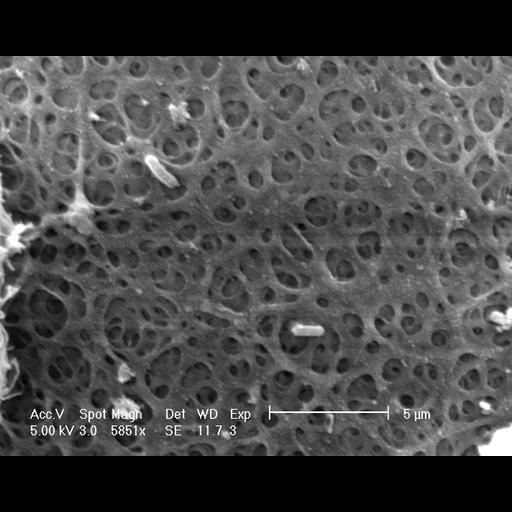
Surface of the zona pellucida surrounding a hamster oocyte. This scanning electron micrograph shows the zona pellicida around a hamster oocyte at high magnification. The zona pellicida has a complex weave and is comprised of three glycoproteins, ZP1, ZP2 and ZP3. The sample was fixed using glutaraldehyde and osmium tetroxide, dehydrated in ethanol, critically point dried, coated with gold, and examined in a Phillips XL30 FEG scanning electron microscope. Magnification: 5851x.

Biological Sources
NCBI Organism Classification
Mesocricetus auratus
Cell Type
oocyte
Cellular Component
zona pellucida
Attribution
Names
Prue Talbot
Citation
Digital Object Identifier (DOI)
doi:10.7295/W9CIL12624
Archival Resource Key (ARK)
ark:/b7295/w9cil12624
Imaging
Image Type
recorded image
Image Mode
scanning electron microscopy (SEM)
Parameters Imaged
elastic scattering of electrons
Source of Contrast
differences in deposition of metal shadow
Visualization Methods
gold
Processing History
unprocessed raw data
Data Qualifiers
raw, unprocessed data
Dimensions
Spatial Axis Image Size Pixel Size
X 619px 0.035µm
Y 484px 0.035µm