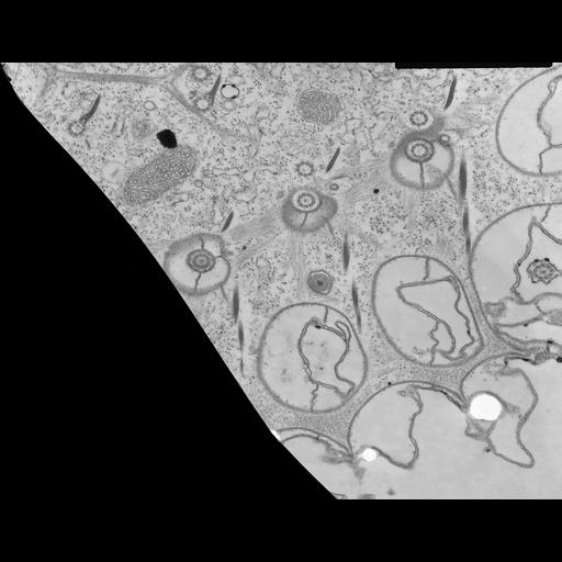
This high resolution EM features the striated bands that link the membranes under the surface depressions of the cortex. The bands insert into the epiplasm, and in this case, they separate to pass around the parasomal sacs which lie to the right of a row of basal body/cilium complexes. The bands have relatively electron transparent zones next to the epiplasm that lie next to more opaque zones which are in turn separated by a central medium electron transparent zone. This differential banding suggests an overlap of filaments in the two more opaque zones, but there is no evidence of a muscle-like actomyosin system. These bands probably provide tension and keep the cortical ridges from flattening. If contractile they could account for the cell’s ability to alter its shape when swimming through constricted regions. For details on the striated bands in Paramecium see Allen, J. Cell Biol. 49:1-20, 1971 and Allen, Aihara and Fok, J. Euk . Microbiol. 45:202-209, 1998. This image is adapted with permission from Fig. 14 in J. Cell Biol. 49:1-20, 1971. TEM taken on 12/18/68 by R. Allen with Philips 300 operating at 60kV. Neg. 16,000X. The raw film was scanned with an Epson Perfection V750 Pro. This image is best used for quantitative analysis. Standard glutaraldehyde fixation followed by osmium tetroxide, dehydrated in alcohol and embedded in an epoxy resin. Microtome sections prepared at approximately 75nm thickness. Additional information available at (http://www5.pbrc.hawaii.edu/allen/).
| Spatial Axis | Image Size | Pixel Size |
|---|---|---|
| X | 6124px | 0.94nm |
| Y | 4768px | 0.94nm |