Licensing
Attribution No Derivatives:This image is licensed under a Creative Commons Attribution, No Derivatives License. View License Deed | View Legal Code
Comments:
Powered by Pnyxe
Description
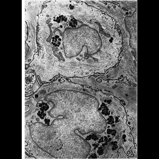
Figure 300 from Chapter 11 (Melanin Pigment) of 'The Cell, 2nd Ed.' by Don W. Fawcett M.D. Conjunctival epithelium from the limbal region of the eye of the Macaca mulatta. Clusters of melanosomes donated from melanocytes (not present in this micrograph) are contained within the epithelium. Intercellular spaces are filled with horseradish peroxidase, injected intravenously, permeating the epithelium and outlining the cells. Image by Giuseppina Raviola. A PDF copy of the accompanying chapter is available on the ASCB's BioEDUCATE website.
Biological Sources
- NCBI Organism Classification
- Macaca mulatta
- (rhesus monkey)
- Cell Type
- epithelial cell
- Cellular Component
- melanosome
Biological Context
- Biological Process
- pigmentation
Attribution
- Names
- Don W. Fawcett
- Giuseppina Raviola
- Published
- ISBN 0721635849
- Link
- http://www.ascb.org/bioeducate/Fawcett...
Grouping
This image is part of a group.Imaging
- Image Type
- transmission electron microscopy (TEM)
- Imaging Mode
- illumination by electrons
- detection of electrons
- Parameters Imaged
- electron density
- Source of Contrast
- stain with broad specificity
- Visualization Methods
- osmium tetroxide
- Data Qualifiers
- processed data
Sample Preparation
- Methods
- crosslinking-fixative fixed tissue
- tissue in plastic embedment
- Relation To Intact Cell
- microtome-sectioned tissue
Dimensions
| Spatial Axis | Image Size | Pixel Size |
|---|---|---|
| X | 910px | —— |
| Y | 1252px | —— |
*CIL – Cell Image Library accession number. Please use this to reference an image.