Alternate header for print version


Licensing
Attribution Non-Commercial; No Derivatives:This image is licensed under a Creative Commons Attribution, Non-Commercial, No Derivatives License. View License Deed | View Legal Code
tweet  
*CIL – Cell Image Library accession number. Please use this to reference an image.

CIL:11465*  Cite 
Description

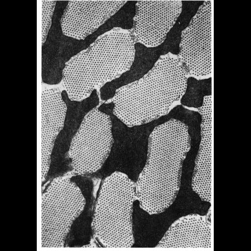
The high wing beat frequency in insects is intense muscle work, and dependent on a ready supply of ATP. This micrograph of flight muscle from the moth, Phytometra gamma shows the physical relationship between mitochondria and myofibrils. Discrete myofibrils of uniform size are surrounded by large mitochondria (sarcosomes), which may exhibit unusual shapes. In this species, a very dense mitochondrial matrix obscures the numerous closely packed cristae. From J. Auber, C.R. Acad. Sci. (Paris) 264: 621-4, 1967, and reprinted here as Figure 261 from Chapter 7 (Mitochondria) of 'The Cell, 2nd Ed.' by Don W. Fawcett M.D. A PDF copy of the accompanying chapter is available on the ASCB’s BioEDUCATE website.

Biological Sources
NCBI Organism Classification
Lepidoptera
Cell Type
flight muscle cell
Cellular Component
mitochondrion
mitochondrial crista
myofibril
Biological Context
Biological Process
cellular respiration
Attribution
Names
J. Auber
Published
ISBN 0721635849
Pubmed
PMID:4962098
Link
Fawcett's "The Cell" 2nd Ed.
Citation
Digital Object Identifier (DOI)
doi:10.7295/W9CIL11465
Archival Resource Key (ARK)
ark:/b7295/w9cil11465
Grouping This image is part of a group.
Imaging
Image Type
transmission electron microscopy (TEM)
Image Mode
illumination by electrons
detection of electrons
Parameters Imaged
electron density
Source of Contrast
stain with broad specificity
Data Qualifiers
processed data
Sample Preparation
Methods
crosslinking-fixative fixed tissue
embedded tissue
Relation To Intact Cell
microtome-sectioned tissue
Dimensions
Spatial Axis Image Size Pixel Size
X 906px ——
Y 1284px ——