Alternate header for print version


Licensing
Attribution Non-Commercial; No Derivatives:This image is licensed under a Creative Commons Attribution, Non-Commercial, No Derivatives License. View License Deed | View Legal Code
tweet  
*CIL – Cell Image Library accession number. Please use this to reference an image.

CIL:11458*  Cite 
Description

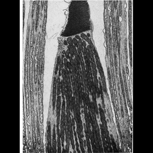
The inner segment of photoreceptors in the retina is packed with long, thin mitochondria that contain curved cristae. This image by Toichiro Kuwabara is of a photoreceptor from human retina, and appears as Figure 257 from Chapter 7 (Mitochondria) of 'The Cell, 2nd Ed.' by Don W. Fawcett M.D. A PDF copy of the accompanying chapter is available on the ASCB’s BioEDUCATE website.

Biological Sources
NCBI Organism Classification
Homo sapiens
Cell Type
eye photoreceptor cell
Cellular Component
mitochondrion
mitochondrial crista
photoreceptor inner segment
Biological Context
Biological Process
cellular respiration
Attribution
Names
Toichiro Kuwabara
Published
ISBN 0721635849
Link
Fawcett's "The Cell" 2nd Ed.
Citation
Digital Object Identifier (DOI)
doi:10.7295/W9CIL11458
Archival Resource Key (ARK)
ark:/b7295/w9cil11458
Grouping This image is part of a group.
Imaging
Image Type
transmission electron microscopy (TEM)
Image Mode
illumination by electrons
detection of electrons
Parameters Imaged
electron density
Source of Contrast
stain with broad specificity
Data Qualifiers
processed data
Sample Preparation
Methods
crosslinking-fixative fixed tissue
embedded tissue
Relation To Intact Cell
microtome-sectioned tissue
Dimensions
Spatial Axis Image Size Pixel Size
X 920px ——
Y 1240px ——