Alternate header for print version


Licensing
Attribution Non-Commercial; No Derivatives:This image is licensed under a Creative Commons Attribution, Non-Commercial, No Derivatives License. View License Deed | View Legal Code
tweet  
*CIL – Cell Image Library accession number. Please use this to reference an image.

CIL:11435*  Cite 
Description

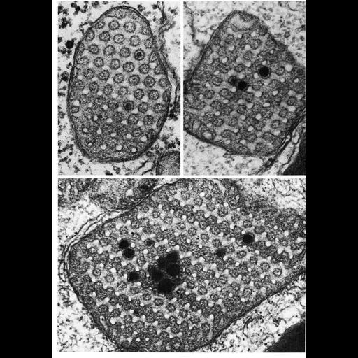
A lattice arrangement of mitochondria is rare in vertebrate cells, but can be observed here in mature Sertoli cells of the Xenopus testes. Multiple parallel fenestrated layers of cristae are interconnected by regularly arranged tubular elements, and shown here in different planes of section. Image from Kalt (1974) Anat. Record. 182:53-60. Figure 244 from Chapter 7 (Mitochondria) of 'The Cell, 2nd Ed.' by Don W. Fawcett M.D. A PDF copy of the accompanying chapter is available on the ASCB’s BioEDUCATE website.

Biological Sources
NCBI Organism Classification
Xenopus
Cell Type
Sertoli cell
Cellular Component
mitochondrion
mitochondrial crista
mitochondrial outer membrane
mitochondrial inner membrane
Biological Context
Biological Process
cellular respiration
Attribution
Names
Marvin Kalt
Published
ISBN 0721635849
Pubmed
PMID: 1155790
Link
Fawcett's "The Cell" 2nd Ed.
Citation
Digital Object Identifier (DOI)
doi:10.7295/W9CIL11435
Archival Resource Key (ARK)
ark:/b7295/w9cil11435
Grouping This image is part of a group.
Imaging
Image Type
transmission electron microscopy (TEM)
Image Mode
illumination by electrons
detection of electrons
Parameters Imaged
electron density
Source of Contrast
stain with broad specificity
Data Qualifiers
processed data
Sample Preparation
Methods
crosslinking-fixative fixed tissue
embedded tissue
Relation To Intact Cell
microtome-sectioned tissue
Dimensions
Spatial Axis Image Size Pixel Size
X 910px ——
Y 1280px ——