Alternate header for print version


Licensing
Attribution Non-Commercial; No Derivatives:This image is licensed under a Creative Commons Attribution, Non-Commercial, No Derivatives License. View License Deed | View Legal Code
tweet  
*CIL – Cell Image Library accession number. Please use this to reference an image.

CIL:11408*  Cite 
Description

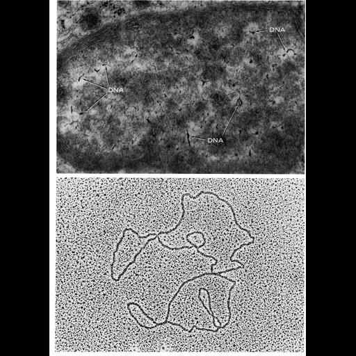
Figures 228 (upper) and 229 (lower) from Chapter 7 (Mitochondria) of 'The Cell, 2nd Ed.' by Don W. Fawcett M.D. A mitochondrion from rat adrenal tissue grown in vitro shows an unusually high number of DNA filaments (upper). Lower, DNA from disrupted Xenopus laevis oocyte mitochondria. Images by Arvi Kahri (upper) and Igor David (lower). A PDF copy of the accompanying chapter is available on the ASCB’s BioEDUCATE website.

Biological Sources
NCBI Organism Classification
Rattus
Xenopus laevis
Cell Type
oocyte
Cellular Component
mitochondrion
mitochondrial chromosome
Biological Context
Biological Process
mitochondrial DNA replication
Attribution
Names
Arvi Kahri
Igor David
Published
ISBN 0721635849
Link
Fawcett's "The Cell" 2nd Ed.
Citation
Digital Object Identifier (DOI)
doi:10.7295/W9CIL11408
Archival Resource Key (ARK)
ark:/b7295/w9cil11408
Grouping This image is part of a group.
Imaging
Image Type
transmission electron microscopy (TEM)
Image Mode
illumination by electrons
detection of electrons
Parameters Imaged
electron density
Source of Contrast
stain with broad specificity
Data Qualifiers
processed data
Sample Preparation
Methods
crosslinking-fixative fixed tissue
embedded tissue
Relation To Intact Cell
isolated subcellular component
Dimensions
Spatial Axis Image Size Pixel Size
X 918px ——
Y 1272px ——