Alternate header for print version


Licensing
Attribution Non-Commercial; No Derivatives:This image is licensed under a Creative Commons Attribution, Non-Commercial, No Derivatives License. View License Deed | View Legal Code
tweet  
*CIL – Cell Image Library accession number. Please use this to reference an image.

CIL:11405*  Cite 
Description

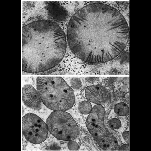
Figures 224 (upper) and 225 (lower) from Chapter 7 (Mitochondria) of 'The Cell, 2nd Ed.' by Don W. Fawcett M.D. Mitochondrial granules range in size from 25 - 120 nm and vary by tissue and physiological state. Shown here in mitochondria from bat liver (upper) and from the guinea pig proximal convoluted tubule. Upper image by Susumu Ito. A PDF copy of the accompanying chapter is available on the ASCB’s BioEDUCATE website.

Biological Context
Biological Process
cellular respiration
Attribution
Names
Don W. Fawcett
Susumu Ito
Published
ISBN 0721635849
Link
Fawcett's "The Cell" 2nd Ed.
Citation
Digital Object Identifier (DOI)
doi:10.7295/W9CIL11405
Archival Resource Key (ARK)
ark:/b7295/w9cil11405
Grouping This image is part of a group.
Imaging
Image Type
transmission electron microscopy (TEM)
Image Mode
illumination by electrons
detection of electrons
Parameters Imaged
electron density
Source of Contrast
stain with broad specificity
Visualization Methods
osmium tetroxide
Data Qualifiers
processed data
Sample Preparation
Methods
crosslinking-fixative fixed tissue
embedded tissue
Relation To Intact Cell
microtome-sectioned tissue
Dimensions
Spatial Axis Image Size Pixel Size
X 906px ——
Y 1264px ——