Alternate header for print version


Licensing
Attribution Non-Commercial; No Derivatives:This image is licensed under a Creative Commons Attribution, Non-Commercial, No Derivatives License. View License Deed | View Legal Code
tweet  
*CIL – Cell Image Library accession number. Please use this to reference an image.

CIL:11232*  Cite 
Description

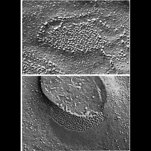
Gap junctions in ciliary epithelium under conditions of oxygenation (upper) and anoxia (lower), in tissue prepared by ultrarapid freezing using liquid helium. Under oxygenated conditions, the connexons are spaced apart, and show no ordered arrangement. Under anoxia, the gap junction has hexagonally packed connexons. Images by Elio Raviola appear as Figures 100 (upper) and 101 (lower) from Chapter 3 (Junctional Specializations) of 'The Cell, 2nd Ed.' by Don W. Fawcett M.D. A PDF copy of the accompanying chapter is available on the ASCB's BioEDUCATE website.

Biological Context
Biological Process
cell communication
cell-cell junction organization
Attribution
Names
Elio Raviola
Published
ISBN 0721635849
Link
The Cell, 2nd Ed. by D.W. Fawcett
Citation
Digital Object Identifier (DOI)
doi:10.7295/W9CIL11232
Archival Resource Key (ARK)
ark:/b7295/w9cil11232
Grouping This image is part of a group.
Imaging
Image Type
transmission electron microscopy (TEM)
Image Mode
illumination by electrons
detection of electrons
Parameters Imaged
electron density
Source of Contrast
differences in deposition of metal shadow
Visualization Methods
shadowing and plating
Data Qualifiers
processed data
Sample Preparation
Methods
freeze_fracture/freeze_etch
Relation To Intact Cell
isolated subcellular component
Dimensions
Spatial Axis Image Size Pixel Size
X 906px ——
Y 1248px ——