Alternate header for print version


Licensing
Attribution Non-Commercial; No Derivatives:This image is licensed under a Creative Commons Attribution, Non-Commercial, No Derivatives License. View License Deed | View Legal Code
tweet  
*CIL – Cell Image Library accession number. Please use this to reference an image.

CIL:11221*  Cite 
Description

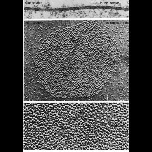
Typical organization of gap junctions as seen in thin sections (upper) and replicas of freeze-fractured cell membranes (middle and lower). Upper, gap junction from the saccular macula of a goldfish; middle, freeze-fracture preparation of a gap junction from a granulosa cell of the rat ovary; lower, freeze-fracture replica of a gap junction from the saccular macula of the goldfish. Images by K. Hama (upper, Fig. 93, lower, Fig. 95), and D. Albertini (middle, Fig. 94) from Chapter 3 (Junctional Specializations) of 'The Cell, 2nd Ed.' by Don W. Fawcett M.D. A PDF copy of the accompanying chapter is available on the ASCB's BioEDUCATE website.

Biological Sources
NCBI Organism Classification
Carassius auratus
Rattus
Cell Type
sensory epithelial cell
granulosa cell
Cellular Component
gap junction
connexon complex
plasma membrane
Biological Context
Biological Process
cell communication
cell-cell junction organization
Attribution
Names
Kiyoshi Hama
David Albertini
Published
ISBN 0721635849
Link
The Cell, 2nd Ed. by D.W. Fawcett
Citation
Digital Object Identifier (DOI)
doi:10.7295/W9CIL11221
Archival Resource Key (ARK)
ark:/b7295/w9cil11221
Grouping This image is part of a group.
Dimensions
Spatial Axis Image Size Pixel Size
X 887px ——
Y 1268px ——