Alternate header for print version


Licensing
Attribution Non-Commercial; No Derivatives:This image is licensed under a Creative Commons Attribution, Non-Commercial, No Derivatives License. View License Deed | View Legal Code
tweet  
*CIL – Cell Image Library accession number. Please use this to reference an image.

CIL:11215*  Cite 
Description

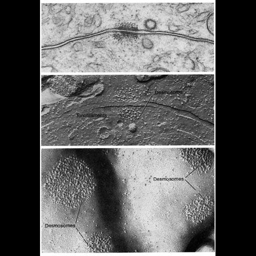
Images of desmosomes from conventional EM and freeze fracture preparations show transmembrane filaments that link two hemidesmosomes. Upper: section of a cell boundary in the ciliary epithelium of the eye; middle, cross fracture across the boundary between two ciliary epithelial cells; lower, replica of a portion of the plasma membrane of an epidermal cell from newborn mouse. Figures 90 (upper), 91 (middle) both courtesy of Giuseppina Raviola, and 92 (lower, from Peter Elias and Daniel Friend) from Chapter 3 (Junctional Specializations) of 'The Cell, 2nd Ed.' by Don W. Fawcett M.D. A PDF copy of the accompanying chapter is available on the ASCB's BioEDUCATE website.

Biological Sources
NCBI Organism Classification
Mus musculus
Cell Type
non-pigmented ciliary epithelial cell
epidermal cell
Cellular Component
desmosome
intermediate filament
keratin filament
plasma membrane
Biological Context
Biological Process
cell adhesion
cell-cell junction organization
Attribution
Names
Giuseppina Raviola
Peter Elias
Daniel Friend
Published
ISBN 0721635849
Link
The Cell, 2nd Ed. by D.W. Fawcett
Citation
Digital Object Identifier (DOI)
doi:10.7295/W9CIL11215
Archival Resource Key (ARK)
ark:/b7295/w9cil11215
Grouping This image is part of a group.
Dimensions
Spatial Axis Image Size Pixel Size
X 898px ——
Y 1307px ——