Alternate header for print version


Licensing
Attribution Non-Commercial; No Derivatives:This image is licensed under a Creative Commons Attribution, Non-Commercial, No Derivatives License. View License Deed | View Legal Code
tweet  
*CIL – Cell Image Library accession number. Please use this to reference an image.

CIL:11150*  Cite 
Description

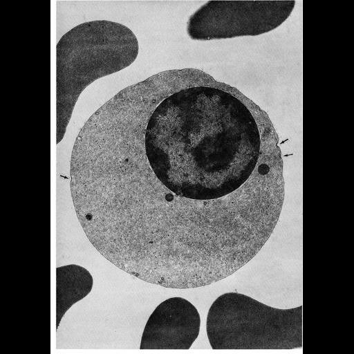
This transmission electron micrograph of a section through a late orthochromatic erythroblast (normoblast) from human bone marrow shows shallow depressions along the membrane surface, indicated by arrows. The thickened membrane at these sites suggests the formation of coated vesicles during micropinocytosis in these erythropoietic cells. The nucleus contains much compact heterochromatin as transcription ceases and is followed by it's ejection.

Technical Details

Figure 53 from Chapter 2 (Specializations of the Free Surface) of 'The Cell, 2nd Ed.' by Don W. Fawcett M.D. A PDF copy of the accompanying chapter is available on the ASCB’s BioEDUCATE website.

Biological Sources
NCBI Organism Classification
Homo sapiens
Cell Type
orthochromatic erythroblast
Cellular Component
coated vesicle
plasma membrane
nucleus
Biological Context
Biological Process
pinocytosis
nucleus organization
Attribution
Names
Don W. Fawcett
Published
ISBN 0721635849
Link
Fawcett's "The Cell, 2nd Ed."
Citation
Digital Object Identifier (DOI)
doi:10.7295/W9CIL11150
Archival Resource Key (ARK)
ark:/b7295/w9cil11150
Grouping This image is part of a group.
Imaging
Image Type
transmission electron microscopy (TEM)
Image Mode
illumination by electrons
detection of electrons
Parameters Imaged
electron density
Source of Contrast
stain with broad specificity
Visualization Methods
osmium tetroxide
Data Qualifiers
processed data
Dimensions
Spatial Axis Image Size Pixel Size
X 914px ——
Y 1280px ——