Alternate header for print version


Licensing
Attribution Non-Commercial; No Derivatives:This image is licensed under a Creative Commons Attribution, Non-Commercial, No Derivatives License. View License Deed | View Legal Code
tweet  
*CIL – Cell Image Library accession number. Please use this to reference an image.

CIL:11137*  Cite 
Description

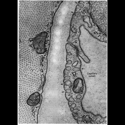
Capillary endothelial cells from mammalian cardiac muscle caught in the act of fluid-phase micropinocytosis. Arrows indicate multiple sites where invaginaton along the membrane suggests vesicles forming during an endocytic event. Figure 52 from Chapter 2 (Specializations of the Free Surface) of 'The Cell, 2nd Ed.' by Don W. Fawcett M.D. A PDF copy of the accompanying chapter is available on the ASCB’s BioEDUCATE website.

Biological Sources
NCBI Organism Classification
Mammalia
Cell Type
endothelial cell
Cellular Component
vesicle
plasma membrane
caveola
Biological Context
Biological Process
pinocytosis
endocytosis
Attribution
Names
Don W. Fawcett
Published
ISBN 0721635849
Link
Fawcett's "The Cell, 2nd Ed."
Citation
Digital Object Identifier (DOI)
doi:10.7295/W9CIL11137
Archival Resource Key (ARK)
ark:/b7295/w9cil11137
Grouping This image is part of a group.
Imaging
Image Type
transmission electron microscopy (TEM)
Image Mode
illumination by electrons
detection of electrons
Parameters Imaged
electron density
Source of Contrast
stain with broad specificity
Visualization Methods
osmium tetroxide
Data Qualifiers
processed data
Dimensions
Spatial Axis Image Size Pixel Size
X 914px ——
Y 1272px ——