Alternate header for print version


Licensing
Attribution Non-Commercial; No Derivatives:This image is licensed under a Creative Commons Attribution, Non-Commercial, No Derivatives License. View License Deed | View Legal Code
tweet  
*CIL – Cell Image Library accession number. Please use this to reference an image.

CIL:11131*  Cite 
Description

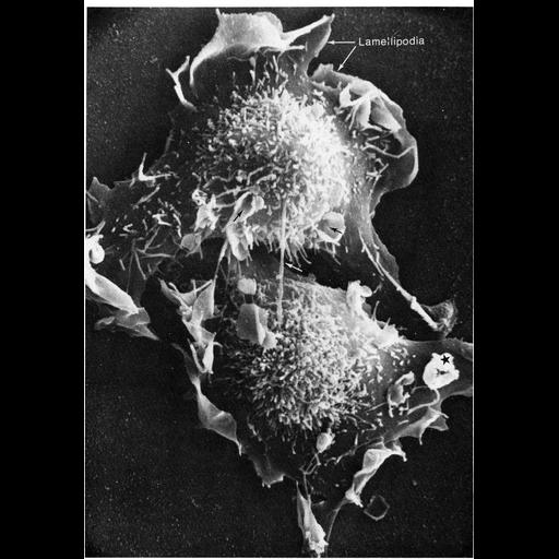
The final stages of cell division is captured in the scanning electron micrograph. Two daughter cells are still interconnected by slender strands of membrane (white arrow at center of image). Lamellipodia extend from the periphery of each new cell as they begin to spread on the substrate, and microvilli stud the surface over the nucleus. The funnel-like cones (black arrows) are candidate sites for pinocytosis. Image contributed by Keith Porter first appeared in Porter and Bonneville, The Fine Structure of Cells and Tissues, Philadelphia: Lea and Febiger, 1973, and reprinted with permission as Figure 49 from Chapter 2 (Specializations of the Free Surface) of 'The Cell, 2nd Ed.' by Don W. Fawcett M.D. A PDF copy of the accompanying chapter is available on the ASCB’s BioEDUCATE website.

Biological Sources
Cellular Component
lamellipodium
cell surface
microvillus
Biological Context
Biological Process
mitosis
pinocytosis
Attribution
Names
Keith Porter
Published
ISBN 0721635849
Link
Fawcett's "The Cell, 2nd Ed."
Citation
Digital Object Identifier (DOI)
doi:10.7295/W9CIL11131
Archival Resource Key (ARK)
ark:/b7295/w9cil11131
Grouping This image is part of a group.
Imaging
Image Type
scanning electron microscopy (SEM)
Image Mode
illumination by electrons
secondary_electron imaging
Parameters Imaged
secondary electron generation
Data Qualifiers
processed data
Sample Preparation
Methods
critical_point dried specimen
Relation To Intact Cell
dispersed cells in vitro
Dimensions
Spatial Axis Image Size Pixel Size
X 894px ——
Y 1280px ——