Alternate header for print version


Licensing
Attribution Non-Commercial; No Derivatives:This image is licensed under a Creative Commons Attribution, Non-Commercial, No Derivatives License. View License Deed | View Legal Code
tweet  
*CIL – Cell Image Library accession number. Please use this to reference an image.

CIL:11111*  Cite 
Description

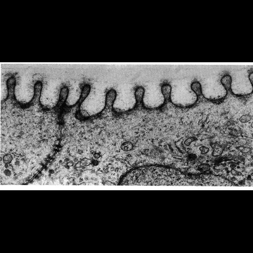
Electron micrograph showing fingerprint like ridges called microplicae on the surface of fish skin epidermis from the neon tetra. These ridges offer mechanical reinforcement against trauma and help hold an abundant mucous layer in place, both protective devices for the skin. Image by Nancy Alexander, Figure 40 from Chapter 2 (Specializations of the Free Surface) of 'The Cell, 2nd Ed.' by Don W. Fawcett M.D. A PDF copy of the accompanying chapter is available on the ASCB’s BioEDUCATE website.

Biological Sources
NCBI Organism Classification
Paracheirodon innesi
Cell Type
epidermal cell
Cellular Component
cell surface
plasma membrane
Biological Context
Biological Process
cell projection organization
plasma membrane organization
Attribution
Names
Nancy Alexander
Published
ISBN 0721635849
Link
Fawcett's "The Cell, 2nd Ed."
Citation
Digital Object Identifier (DOI)
doi:10.7295/W9CIL11111
Archival Resource Key (ARK)
ark:/b7295/w9cil11111
Grouping This image is part of a group.
Imaging
Image Type
transmission electron microscopy (TEM)
Image Mode
illumination by electrons
detection of electrons
Parameters Imaged
electron density
Source of Contrast
stain with broad specificity
Visualization Methods
osmium tetroxide
Data Qualifiers
processed data
Sample Preparation
Methods
crosslinking-fixative fixed tissue
Relation To Intact Cell
microtome-sectioned tissue
Dimensions
Spatial Axis Image Size Pixel Size
X 847px ——
Y 430px ——