Alternate header for print version


Licensing
Attribution Non-Commercial; No Derivatives:This image is licensed under a Creative Commons Attribution, Non-Commercial, No Derivatives License. View License Deed | View Legal Code
tweet  
*CIL – Cell Image Library accession number. Please use this to reference an image.

CIL:11110*  Cite 
Description

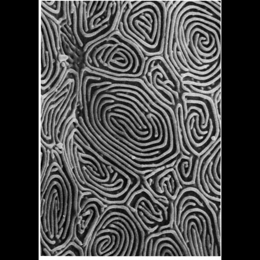
of 'The Cell, 2nd Ed.' by Don W. Fawcett M.D. Scanning electron micrograph of the epidermal surface of the flatfish sole Parophrys detulus shows a labyrinth of surface ridges. Image from Fahrenbach, W., Knutsen, D.D. (1975). J. Invest. Dermatol. 65:39-44, reprinted by permission from Macmillan Publishers Ltd as Figure 41 from Chapter 2 (Specializations Of The Free Surface) of 'The Cell, 2nd Ed.' by Don W. Fawcett M.D. A PDF copy of the accompanying chapter is available on the ASCB's BioEDUCATE website.

Biological Sources
NCBI Organism Classification
Parophrys vetulus
Cell Type
epidermal cell
Cellular Component
cell surface
external side of plasma membrane
Biological Context
Biological Process
plasma membrane organization
cell projection organization
Attribution
Names
Wolf Fahrenbach
Published
ISBN 0721635849
Pubmed
PMID: 1151108
Link
Fawcett's "The Cell, 2nd Ed."
J. Invest. Dermatol. reprint
Citation
Digital Object Identifier (DOI)
doi:10.7295/W9CIL11110
Archival Resource Key (ARK)
ark:/b7295/w9cil11110
Grouping This image is part of a group.
Imaging
Image Type
scanning electron microscopy (SEM)
Image Mode
illumination by electrons
secondary_electron imaging
Parameters Imaged
secondary electron generation
Data Qualifiers
processed data
Sample Preparation
Methods
critical_point dried specimen
Dimensions
Spatial Axis Image Size Pixel Size
X 908px ——
Y 1292px ——