Alternate header for print version


Licensing
Attribution Non-Commercial Share Alike:This image is licensed under a Creative Commons Attribution, Non-Commercial Share Alike License. View License Deed | View Legal Code
tweet  
*CIL – Cell Image Library accession number. Please use this to reference an image.

CIL:11108*  Cite 
Description

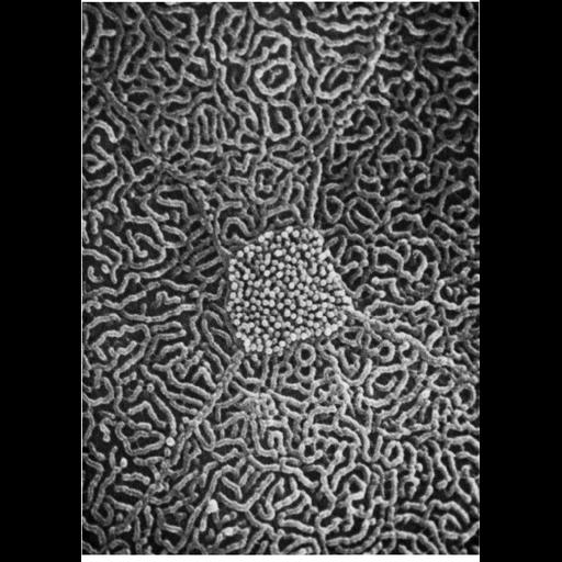
Scanning electron micrograph of the lumenal surface of the toad bladder shows the boundaries of four polygonal squamous epithelial cells surrounding a mitochondrial-rich cell bearing short globular microvilli. Boundaries between cells are demarcated by what appears to be a continuous ridge. Image from Davis, Goodman, & Schuster (1974) J. Cell Biol. 61:544-547 and reprinted with permission as Figure 39 from Chapter 2 (Specializations of the Free Surface) of 'The Cell, 2nd Ed.' by Don W. Fawcett M.D. A PDF copy of the accompanying chapter is available on the ASCB’s BioEDUCATE website.

Biological Sources
NCBI Organism Classification
Bufo
Cell Type
squamous epithelial cell
Cellular Component
cell surface
microvillus
Biological Context
Biological Process
extracellular structure organization
Attribution
Names
Walter L. Davis
D. B. Goodman
Published
ISBN 0721635849
Pubmed
PMID: 4373479
Link
Fawcett's "The Cell, 2nd Ed."
Davis et al. (1974) J. Cell Biol.
OTHER
R. J. Schuster
Citation
Digital Object Identifier (DOI)
doi:10.7295/W9CIL11108
Archival Resource Key (ARK)
ark:/b7295/w9cil11108
Grouping This image is part of a group.
Imaging
Image Type
scanning electron microscopy (SEM)
Image Mode
illumination by electrons
secondary_electron imaging
Parameters Imaged
secondary electron generation
Data Qualifiers
processed data
Sample Preparation
Methods
critical_point dried specimen
Dimensions
Spatial Axis Image Size Pixel Size
X 906px ——
Y 1276px ——