Licensing

Attribution_by_nc_nd
Attribution Non-Commercial; No Derivatives:This image is licensed under a Creative Commons Attribution, Non-Commercial, No Derivatives License. View License Deed | View Legal Code
Comments: Powered by Pnyxe

Description

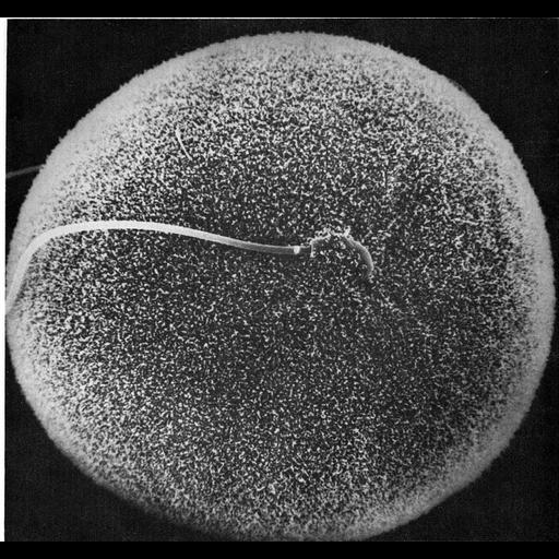
Scanning electron micrograph showing in vitro fertilization of a hamster oocyte. The sperm head, near the center of the field of view, is engulfed by the microvilli on the surface of the oocyte membrane; the sperm tail extends to the left. Image by David Phillips, Figure 32 from Chapter 2 (Specializations of the Free Surface) of 'The Cell, 2nd Ed.' by Don W. Fawcett M.D. . A PDF copy of the accompanying chapter is available on the ASCB's BioEDUCATE website.

Biological Sources

NCBI Organism Classification
Phodopus
(Asiatic Dwarf Hamsters)
Cell Type
oocyte
sperm
Cellular Component
microvillus membrane

Attribution

Name
David Phillips
Published
ISBN 0721635849
Link
Fawcett's "The Cell, 2nd Ed."

Grouping

This image is part of a group.
Data Qualifiers
processed data

Sample Preparation

Methods
critical_point dried specimen
Relation To Intact Cell
dispersed cells in vitro

Dimensions

Spatial Axis Image Size Pixel Size
X 891px ——
Y 836px ——
*CIL – Cell Image Library accession number. Please use this to reference an image.