Alternate header for print version


Licensing
Attribution Non-Commercial; No Derivatives:This image is licensed under a Creative Commons Attribution, Non-Commercial, No Derivatives License. View License Deed | View Legal Code
tweet  
*CIL – Cell Image Library accession number. Please use this to reference an image.

CIL:10944*  Cite 
Description

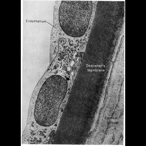
This electron micrograph shows the endothelium and the underlying Descemet's membrane of the human cornea. Descemet's membrane is an unusually thick and structurally specialized example of basal lamina, secreted by the adjacent endothelial cells. The collagen of this layer may exhibit an organized array of evenly spaced nodes connected by radiating fibrils to give a lattice-like appearance. Image by T. Kuwabara, Figure 23 from Chapter 1 (The Cell Surface) of 'The Cell, 2nd Ed.' by Don W. Fawcett M.D. A PDF copy of the accompanying chapter is available on the ASCB's BioEDUCATE website.

Biological Sources
NCBI Organism Classification
Homo sapiens
Cell Type
corneal endothelial cell
Cellular Component
basal lamina
extracellular region part
plasma membrane
Biological Context
Biological Process
cell-substrate adhesion
extracellular structure organization
Attribution
Names
Toichiro Kuwabara
Published
ISBN 0721635849
Link
The Cell, 2nd Ed. by D.W. Fawcett
Citation
Digital Object Identifier (DOI)
doi:10.7295/W9CIL10944
Archival Resource Key (ARK)
ark:/b7295/w9cil10944
Grouping This image is part of a group.
Imaging
Image Type
transmission electron microscopy (TEM)
Image Mode
illumination by electrons
detection of electrons
Parameters Imaged
electron density
Source of Contrast
stain with broad specificity
Visualization Methods
electron-dense stain
Data Qualifiers
processed data
Sample Preparation
Methods
crosslinking-fixative fixed tissue
Relation To Intact Cell
microtome-sectioned tissue
Dimensions
Spatial Axis Image Size Pixel Size
X 906px ——
Y 1268px ——