Alternate header for print version


Licensing
Attribution Non-Commercial; No Derivatives:This image is licensed under a Creative Commons Attribution, Non-Commercial, No Derivatives License. View License Deed | View Legal Code
tweet  
*CIL – Cell Image Library accession number. Please use this to reference an image.

CIL:10937*  Cite 
Description

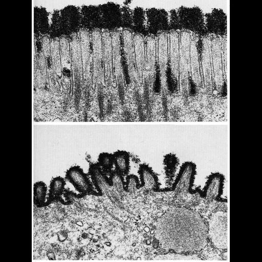
These two micrographs show the outer cell surface of the epithelial cell border in intestine (upper) and stomach (lower) tissue. This surface, called the glycocalyx, is composed of negatively charged acidic polysaccharides, which here, have been stained by binding a polycationic derivative of ferritin. High iron content causes electron scattering, and thus, regions to which the ferritin have bound appear dark in these images. These images, courtesy of Susumu Ito, are Figures 20 (upper) and 21 (lower) from Chapter 1 (The Cell Surface) of 'The Cell, 2nd Ed.' by Don W. Fawcett M.D. A PDF copy of the accompanying chapter is available on the ASCB's BioEDUCATE website.

Biological Context
Biological Process
extracellular structure organization
Attribution
Names
Susumu Ito
Published
ISBN 0721635849
Link
The Cell, 2nd Ed. by D.W. Fawcett
Citation
Digital Object Identifier (DOI)
doi:10.7295/W9CIL10937
Archival Resource Key (ARK)
ark:/b7295/w9cil10937
Grouping This image is part of a group.
Imaging
Image Type
transmission electron microscopy (TEM)
Image Mode
illumination by electrons
detection of electrons
Parameters Imaged
electron density
Source of Contrast
stain with broad specificity
Data Qualifiers
processed data
Sample Preparation
Methods
crosslinking-fixative fixed tissue
Relation To Intact Cell
microtome-sectioned tissue
Dimensions
Spatial Axis Image Size Pixel Size
X 908px ——
Y 1200px ——