Licensing

Attribution_by_nc_nd
Attribution Non-Commercial; No Derivatives:This image is licensed under a Creative Commons Attribution, Non-Commercial, No Derivatives License. View License Deed | View Legal Code
Comments: Powered by Pnyxe

Description

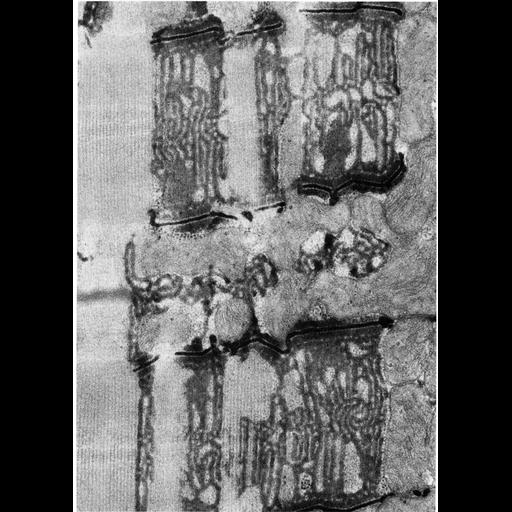
Figure 195 from Chapter 5 (Endoplasmic Reticulum) of 'The Cell, 2nd Ed.' by Don W. Fawcett M.D. Selective staining of tubular invaginations of the sarcolemma in the tibialis anterior of the mouse. Following fixation by glutaraldehyde solution containing calcium, the tissue was post-fixed with osmium ferrocyanide, resulting in staining of the T-tubules and sacrotubules of the reticulum. The adjacent terminal cisternae and longitudinal sarcotubules are lightly stained. Image by Michael Forbes. A PDF copy of the accompanying chapter is available on the ASCB's BioEDUCATE website.

Biological Sources

NCBI Organism Classification
Mus musculus
(house mouse)
Cell Type
skeletal muscle cell
Cellular Component
sarcoplasmic reticulum
myofibril
I band
A band
Z disc
terminal cisterna
sarcomere

Biological Context

Biological Process
striated muscle contraction

Attribution

Name
Michael Forbes
Published
ISBN 0721635849
Link
http://www.ascb.org/bioeducate/Fawcett...

Grouping

This image is part of a group.
Data Qualifiers
processed data

Sample Preparation

Methods
glutaraldehyde fixed tissue
tissue in plastic embedment
Relation To Intact Cell
microtome-sectioned tissue

Dimensions

Spatial Axis Image Size Pixel Size
X 898px ——
Y 1260px ——
*CIL – Cell Image Library accession number. Please use this to reference an image.