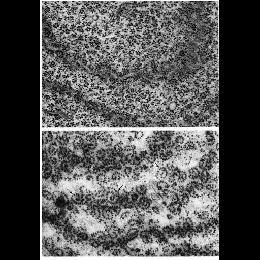
Figures 173 (upper panel) and 174 (lower panel) from Chapter 5 (Endoplasmic Reticulum) of 'The Cell, 2nd Ed.' by Don W. Fawcett M.D. Electron micrographs of endoplasmic reticulum with the section plane parallel to the cisternae, revealing the arrangement of ribsomes along them. The upper panel is from a large neuron and shows numerous free polysomes associated with a Nissl body; arrows capture two regions where the plane is tangential to the cisternae, and shows ribosomes arranged in loops, spirals and rosettes. The lower panel, from a glandular, adrenocortical cell of a human fetus at 27 weeks, the section plan is tangential to three cisternae. This view reveals long loops and spirals of multiple ribosomes aligned along a single molecule of messenger RNA. Upper image by Sanford Palay; lower image by Eichi Yamada. A PDF copy of the accompanying chapter is available on the ASCB’s BioEDUCATE website.
| Spatial Axis | Image Size | Pixel Size |
|---|---|---|
| X | 900px | —— |
| Y | 1304px | —— |