Alternate header for print version


Licensing
Public Domain: This image is in the public domain and thus free of any copyright restrictions. However, as is the norm in scientific publishing and as a matter of courtesy, any user should credit the content provider for any public or private use of this image whenever possible. Learn more
tweet  
*CIL – Cell Image Library accession number. Please use this to reference an image.

CIL:10730*  Cite 
Description

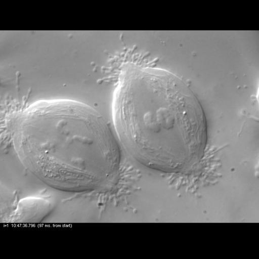
Movie of meiosis I and II in primary spermatocytes of the crane-fly Nephrotoma suturalis. Time-lapse DIC microscopy using a Nikon Microphot SA, equipped for liquid crystal polarized light microscopy (LC-PolScope, CRi, Woburn Massachusetts) 60x/1.4 PlanApo oil immersion objective, 1.4 NA oil imm. condenser, with 2.0x zoom lens. Images captured every 30 sec over 320 min using a QImaging Retigo EXi CCD camera. A movie created from the time series is included in this grouped set.

Biological Sources
NCBI Organism Classification
Nephrotoma
Nephrotoma suturalis
Cell Type
primary spermatocyte
Cellular Component
spindle
nuclear chromosome
Biological Context
Biological Process
male meiosis chromosome segregation
Attribution
Names
James R. LaFountain
Rudolf Oldenbourg
Published
LaFountain JR, Oldenbourg R (2004) Mol Biol Cell 15, 5346-5355.
Pubmed
15385630
Citation
Digital Object Identifier (DOI)
doi:10.7295/W9CIL10730
Archival Resource Key (ARK)
ark:/b7295/w9cil10730
Grouping This image is part of a group.
Imaging
Image Type
charge coupled device (CCD)
Image Mode
differential interference contrast microscopy
Parameters Imaged
UV/visible/IR photons
Source of Contrast
differences in refractive index
Visualization Methods
visualization of contiguous regions
Processing History
unprocessed raw data
Sample Preparation
Methods
living tissue
Relation To Intact Cell
whole mounted tissue
Dimensions
Spatial Axis Image Size Pixel Size
X 678px 87nm
Y 518px 87nm
Time 30 seconds 320