Alternate header for print version


Licensing
Public Domain: This image is in the public domain and thus free of any copyright restrictions. However, as is the norm in scientific publishing and as a matter of courtesy, any user should credit the content provider for any public or private use of this image whenever possible. Learn more
tweet  
*CIL – Cell Image Library accession number. Please use this to reference an image.

CIL:10705*  Cite 
Description

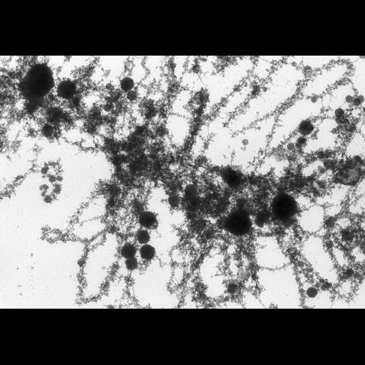
High voltage TEM image of meiotic newt oocyte nuclei showing transcription on looped lampbrush chromosomes. This micrograph, was recorded at a tilt angle of 50 degrees. The image is grouped with one of the same sample area recorded at a 40 degree tilt. A stereo examination of the pair provides an oblique 3D view.

Technical Details

Oocytes were allowed to disperse in 30 mM KCl/NaCl, centrifuged through 2% formaldehyde onto an EM grid, stained with uranyl acetate, and critical point dried. Grids were examined with the Madison 1MeV TEM at 5 KX.

Biological Sources
NCBI Organism Classification
Notophthalmus viridescens
Cell Type
oocyte
Cellular Component
lampbrush chromosomes
Biological Context
Biological Process
Transcription
chromosome organization
RNA metabolic process
Attribution
Names
Hans Ris
Citation
Digital Object Identifier (DOI)
doi:10.7295/W9CIL10705
Archival Resource Key (ARK)
ark:/b7295/w9cil10705
Grouping This image is part of a group.
Imaging
Image Type
film
Image Mode
Parameters Imaged
electron density
Source of Contrast
differences in adsorption or binding of stain
Visualization Methods
uranyl salt
Processing History
unprocessed raw data
Sample Preparation
Methods
formaldehyde fixed tissue
Relation To Intact Cell
isolated subcellular component
Dimensions
Spatial Axis Image Size Pixel Size
X 5984px 2.5nm
Y 4152px 2.5nm