File containing the merged fluorescent-DIC imaging for each time point, where the fluorescent image is a maximum intensity projection of the z series collected at each time point. Each time point is represented as a "slice" in the volume in IMOD format.
File format
MRC
Animation description
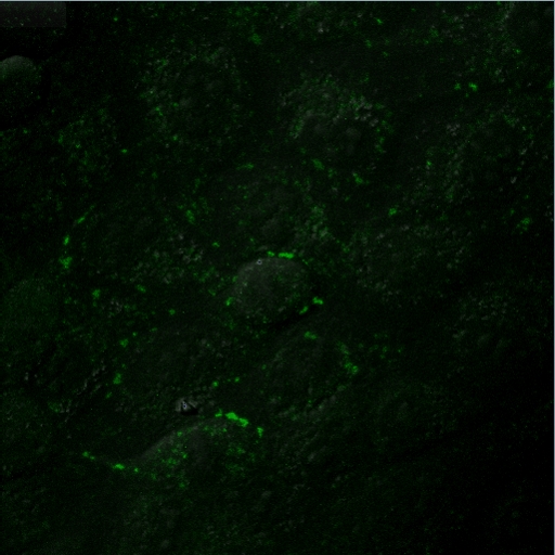
Annnotated time-lapse animation of FlAsH-labeled MDCK cells expressing Cx43-4C309/337 (green) showing their rearrangement and fate during and after mitosis. FlAsH fluorescence excitation z series along with a transmitted DIC image were recorded every 6 min for a total duration of about 5 h. 3D volume reconstructions from confocal image stacks over time are displayed at a rate of two frames per minute. Supplemental movie 2 from Boassa et al. (2010).
Merged fluorescent and DIC image of MDCK cells expressing Cx43-4C309/337 (green)
Full resolution image description
Optical section/time series in Olympus Fluoview .oif format. Each time point represents 11 z slices from the fluorescent channel and a DIC image.
Animation description
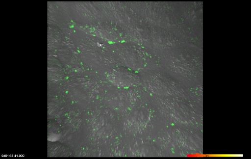
Time-lapse imaging of FlAsH-labeled MDCK cells expressing
Cx43-4C309/337 (green) showing their rearrangement and fate during and after
mitosis. FlAsH fluorescence excitation z series along with a transmitted DIC image were recorded every 6 min for a total duration of about 5 h. 3D volume reconstructions from confocal image stacks over time are displayed at a rate of two frames per minute.
The fate of Connexin43 gap junction protein during mitosis
Description
During the cell cycle, gap junction communication, morphology and distribution of connexin43 (Cx43)-containing structures change dramatically. As cells round up in mitosis, the majority of Cx43 labeling is intracellular and intercellular coupling is reduced. We are interested in investigating Cx43 distributions during mitosis in exogenous expressing cells using in vivo time-lapse imaging.
Funding agency
NIH GM072881 and GM065937 awarded to Gina Sosinky, NIH GM55632 awarded to Paul Lampe
Leader(s)
Daniela Boassa
Collaborator(s)
Gina Sosinky
Paul Lampe and Joell Solan
Start date
unspecified
End date
unspecified
Experiment
Experiment ID
7789
Title
Live cell imaging of rCx43-309/337-4C stable cell line
Purpose
To test whether the 'old' Cx43 proteins labeled before mitosis could be re-utilized to form new gap junctions upon the mitotic phase exit, we performed time-lapse imaging of green FlAsH-labeled rCx43-309/337 4C and followed their fate during and after completion of mitosis.
Experimenter(s)
Daniela Boassa
Microscopy product
Microscopy product ID
7796
Instrument
Olympus Fluoview 1000
Microscopy type
LASER SCANNING CONFOCAL
Product type
TIME SERIES
Image basename
Supplemental movie 2
Spatial Axis
Image Size
Pixel Size
X
512px
0.276 um/pixels
Y
512px
0.276 um/pixels
Subject
Species
Dog
Scientific name
Canis familiaris
Strain
NA
Group by
NA
Age class
NA
Tissue section
Anatomical location
NA
Specimen description
Organ
kidney
Structure
gap junction
Cell type
MDCK cell
Imaging parameters
Type
Light microscopy product
Immersion medium
oil
Mounting medium
Opti-MEM
Lens
Olympus PlanApo 60X oil
Lens magnification
X
Numerical aperture
1.42
Notes
Medium: Opti-MEM supplemented with 5% FBS covered with a glass coverslip.
Specimen preparation
Protocol used
MDCK cells stably expressing two internal 4C domains in the C-terminus (FLNCCPGCCME)-tagged Cx43 (Cx43-4C309/337) were labeled for 1 h at 37 degrees C with 180 nM FlAsH-EDT2/12.5 uM EDT in Hanks balanced salt saline (HBSS). Free and non-specifically bound FlAsH was removed by washing with 2,3-dimercapto-1-propanol (BAL, 500 uM, 20min at 37 degrees C in HBSS). Time-lapse imaging was conducted using an Olympus FluoView1000 confocal microscope equipped with a temperature controlled chamber (at 37 degrees C) and a 60X 1.42 NA objective. Medium: Opti-MEM supplemented with 5% FBS covered with a glass coverslip.
Imaging product type
Type
Optical section
Description
For Movie S2 both the FlAsH fluorescence and the differential interference contrast (DIC) images were collected, 515 nm excitation/530¿630 nm emission.
Images were recorded every 6 min for a total duration of about 5 h,
0.276 ¿m/pixel, image size 512 × 512 pixels in 11 z stacks (step size
1 ¿m), 12 bits/pixel, sampling speed 2 ¿s/pixel.
Z resolution
1 um
Citation Information
Daniela Boassa, Gina Sosinky, Paul Lampe and Joell Solan CCDB:7796, Canis familiaris, gap junction, MDCK cell. CIL. Dataset. https://doi.org/doi:10.7295/W9CCDB7796