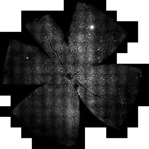
Mosaic reconstruction imaged using spinning disk confocal microscopy of a flat mount of retina showing retinal ganglion cells retrogradely labeled via fluorogold injection into the superior colliculus in an astrocyte specific Nf1 heterozygote (Nf1 -/+) conditional knock out mouse. Details may be found in Kim et al., Neuroscience. 2010 Sep 29;170(1):178-88.
Full resolution image description
Assembled mosaic in tiff format of a flat mounted retina with retinal ganglion cells retrogradely labeled via fluorogold: Nf1_CKOH.tif
Characterization of optic gliomas from neurofibromatosis-1 (Nf1) genetically-engineered mice
Description
Use of large scale imaging of the optic nerve and chiasm to characterize development of optic gliomas from neurofibromatosis-1 (Nf1) genetically-engineered mice
Funding agency
NCI and NCRR
Leader(s)
Keun-Young Kim
Collaborator(s)
David Gutmann
Eric Bushong
Mark Ellisman
Da-Yong Lee
Start date
09-01-2009
End date
unspecified
Experiment
Experiment ID
7723
Title
Large scale mosaic imaging with retrograde labeling of retinal ganglion cells
Purpose
The purpose of this study was to investigate the progression of changes in retinal ganglion cells and optic nerve glia in neurofibromatosis-1 (NF1) genetically-engineered mice with optic glioma. RGC were counted in whole retina preparations using high-resolution, mosaic confocal microscopy following their delineation by retrograde FluoroGold labeling. Then, we found reduced RGC numbers in Nf1+/¿GFAPCKO mice, supporting a model in which the combination of optic nerve Nf1 heterozygosity and glial cell Nf1 loss results in disrupted axonal-glial relationships, subsequently culminating in the degeneration of optic nerve axons and loss of their parent RGC neurons.
Experimenter(s)
Keun-Young Kim
Microscopy product
Microscopy product ID
7750
Instrument
Olympus DSU Confocal Microscope
Microscopy type
Spinning Disc Confocal Microscopy
Product type
MOSAIC
Image basename
Nf1_CKOH
Subject
Species
mouse
Scientific name
Mus musculus
Strain
C57Bl/6 Nf1+/- GFAP CKO
Group by
Injection of tracer
Treatment
Both mutant and wild-type mice were injected with fluorogold tracer bilaterally into the superior colliculi. Please refer to the subject description for which images belong to which type of animal.
Retrograde Labeling of Retinal Ganglion Cells
One week prior to euthanasia, FluoroGold (1 ul/injection; Fluorochrome Inc., Englewood, CO) diluted in saline was microinjected bilaterally into the superior colliculi of anesthetized mice with a mixture of ketamine (Fort Dodge Animal Health) and xylazine (Vedeco Inc.) in a stereotactic apparatus, as previously described (Kim et al., 2004). Fluoro-Gold is taken up by the axon terminals of RGC neurons and transported retrogradely to the cell bodies in the retina (Selles-Navarro et al., 1996). The FluoroGold in the RGC neurons persists for at least 3 weeks without significant fading or leakage (Dong et al., 1996). Mosaic image arrays from retinal flat-mounts (n=6 retinal flat-mounts/mice/group) were captured under an Olympus spinning disk confocal microscope (Olympus America Inc., Center Valley, PA) equipped with a high-precision closed loop XY stage and closed loop Z control with commercial mosaic acquisition software from MicroBrightField (MBF Bioscience Inc., Williston, VT) modified by us (Chow et al., 2006, Price et al., 2006). The microscope was equipped with a high-resolution CCD camera for high-speed mosaic acquisition. Images were stored in Photoshop files (Adobe Systems Inc., San Jose, CA).
Age
9 months
Age class
Adult
Tissue section
Anatomical location
eye
Thickness
160 µm
Specimen description
Organ
eye
System
central nervous system
Structure
cell body
Tissue
retina
Cell type
retinal ganglion cell
Imaging parameters
Type
Light microscopy product
Immersion medium
air
Mounting medium
gelvatol
Lens
Olympus 20x dry
Numerical aperture
.75
Notes
christine
Imaging product type
Type
Mosaic
Description
FG labled RGC in flatmounted retina
Citation Information
Keun-Young Kim, David Gutmann, Eric Bushong, Mark Ellisman, Da-Yong Lee (2009) CCDB:7750, Mus musculus, cell body, retinal ganglion cell. CIL. Dataset. https://doi.org/doi:10.7295/W9CCDB7750