Computed slices through a double tilt electron tomographic reconstruction of synaptic terminals in rat cultured cortical neurons expressing alpha synuclein-MiniSOG and processed for photo-oxidation. Extensive stained membranous elements are observed in the pre-synaptic terminal Data corresponds to Fig. 4 in Boassa et al. 2013.
Full resolution image description
Computed slices through a double tilt electron tomographic reconstruction of synaptic terminals in rat cultured cortical neurons expressing alpha synuclein-MiniSOG and processed for photo-oxidation. Extensive stained membranous elements are observed in the pre-synaptic terminal Data corresponds to Fig. 4 (Tomogram of cultured neurons expressing AS-MiniSOG processed for photooxidation. Extensive stained membranous elements are observed in the presynaptic terminal.)in Boassa et al. 2013.
Zero degree tilt image of a presynaptic bouton from a cultured neuron expressing alpha synuclein-MiniSOG processed for photo-oxidation. Extensive stained membranous elements are observed in the pre-synaptic terminal. Data shown in Fig. 4B of Boassa et al., 2013. Use the brightness and contrast controls to adjust image.
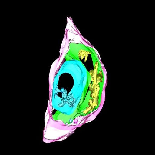
Manual segmentation of plasma membrane and labeled tubero-membranous system in a synaptic terminal
Segmentation file description
Three-dimensional model from EM tomogram of same area as 4B shows plasma membrane (pink) and contiguous membranes in three different colors (blue, green, and yellow).
Molecular and cellular investigations of alpha-synuclein in Parkinson pathogenesis
Description
The goal of this project is to better understand the role of alpha-synuclein in the progression of PD by applying labeling systems developed at NCMIR to track, at multiple resolution scales, the distribution and trafficking of these proteins associated with Parkinson's disease.
Leader(s)
Daniela Boassa
Collaborator(s)
Monica L. Berlanga
Mary Ann Yang
Masako Terada
Junru Hu
Eric A. Bushong
Minju Hwang
Eliezer Masliah
Julia M. George
and Mark H. Ellisman
Start date
unspecified
End date
unspecified
Experiment
Experiment ID
9636
Experiment date
03-08-2011
Title
Distribution of Alpha-Synuclein over-expressed in primary cultured neurons.
Purpose
EM analysis of localization of alpha-synuclein tagged with miniSOG.
Experimenter(s)
Daniela Boassa
Microscopy product
Microscopy product ID
61937
Instrument
JEOL4000EX
Microscopy type
IVEM
Product type
DOUBLE TILT
Image basename
dish1Area4Grid5_1
Subject
Species
Rat
Scientific name
Rattus
Strain
Sprague Dawley
Age
2 days
Age class
Pup
Tissue section
Anatomical location
Brain, cortex
Specimen description
Organ
brain
System
central nervous system
Cell type
Neuron
Imaging parameters
Type
Electron microscopy product
Recording medium
4K CCD
Magification
0
Accelerating voltage
300 KV
Specimen preparation
Protocol used
Primary neurons were prepared from cortex of 2-day-old Harlan Sprague Dawley rats and cultured on poly-d-lysine-coated dishes in Neurobasal-A medium supplemented with B27 and l-glutamine. Before plating, neurons were transfected with the Amaxa Nucleofector protocol and imaged at 17-21 DIV.
Imaging product type
Type
Double tilt
X min range
60 degrees
X max range
-60 degrees
X tilt increment
2 degrees
Y min range
60 degrees
Y max range
-60 degrees
Y tilt increment
2 degrees
Citation Information
Daniela Boassa, Monica L. Berlanga, Mary Ann Yang, Masako Terada, Junru Hu, Eric A. Bushong, Minju Hwang, Eliezer Masliah, Julia M. George, and Mark H. Ellisman CCDB:61937, Rattus, Neuron. CIL. Dataset. https://doi.org/doi:10.7295/W9CCDB61937