Alternate header for print version


Licensing
Public Domain: This image is in the public domain and thus free of any copyright restrictions. However, as is the norm in scientific publishing and as a matter of courtesy, any user should credit the content provider for any public or private use of this image whenever possible. Learn more
tweet  
*CIL – Cell Image Library accession number. Please use this to reference an image.

CIL:9712*  Cite 
Description

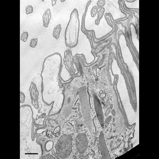
A view near the oral apparatus of Coleps and the attachment site of a microtubule filament bundle, fibrous interconnections, and a rod near a basal body that is out of the section plane in this TEM. Discoidal vesicles probably involved in digestion are aligned along the inside edge of the rod as are the extrusive organelles, the toxicysts. Standard glutaraldehyde fixation followed by osmium tetroxide, dehydrated in alcohol and embedded in an epoxy resin. Microtome sections prepared at approximately 75nm thickness. TEM taken on 5/28/69 by R. Allen with Philips 300 operating at 60kV. Neg. 14,800X. Bar = 0.5µm. A print of the negative was scanned and processed in Photoshop. This image is best used for qualitative analysis. A high resolution image (CIL:9018) is available for quantitative analysis. Additional information available at (http://www5.pbrc.hawaii.edu/allen/).

Biological Sources
NCBI Organism Classification
Coleps hirtus
Cell Type
eukaryotic cell
Eukaryotic Protist
Ciliated Protist
Cell Line
Cambridge Culture Collection, Cambridge, England, catalog # LB1656/1
Cellular Component
cellular ingestive element
plasma membrane
microtubule basal body
cytoskeleton
microtubule cytoskeleton
actin cytoskeleton
Biological Context
Biological Process
digestive system process
microtubule cytoskeleton organization
microfibril organization
Molecular Function
structural molecule activity
Attribution
Names
Richard Allen
Citation
Digital Object Identifier (DOI)
doi:10.7295/W9CIL9712
Archival Resource Key (ARK)
ark:/b7295/w9cil9712
Grouping This image is part of a group.
Imaging
Image Type
transmission electron microscopy (TEM)
illumination by electrons
Image Mode
detection of electrons
film
Parameters Imaged
electron density
Source of Contrast
stain with broad specificity
Visualization Methods
stain with broad specificity
osmium tetroxide
uranyl salt
lead salt
Processing History
recorded image
film
Print of negative scanned to Photoshop.
Data Qualifiers
processed data
suitable for spatial measurements
Dimensions
Spatial Axis Image Size Pixel Size
X 2400px ——
Y 3200px ——