Alternate header for print version


Licensing
Public Domain: This image is in the public domain and thus free of any copyright restrictions. However, as is the norm in scientific publishing and as a matter of courtesy, any user should credit the content provider for any public or private use of this image whenever possible. Learn more
tweet  
*CIL – Cell Image Library accession number. Please use this to reference an image.

CIL:9704*  Cite 
Description

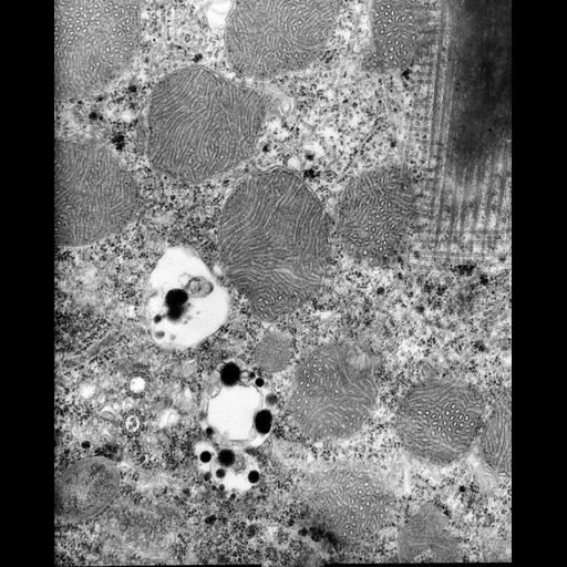
A high resolution image of a dikinetid of Euplotes shows the transverse microtubular ribbon as well as a postciliary ribbon extending from the posterior basal body. Vesicles containing electron opaque bodies are typically found next to basal bodies. A peroxisome is also present. In this section a pattern of cross-hatched microtubules lies immediately under the cell surface membranes. Standard glutaraldehyde fixation followed by osmium tetroxide, dehydrated in alcohol and embedded in an epoxy resin. Microtome sections prepared at approximately 75nm thickness. TEM taken on 8/3/67 by R. Allen with Philips 200 operating at 60kV. Neg. 19,200X. The raw negative was scanned with an Epson Perfection V750 Pro. This image is best used for quantitative analysis. Additional information is available at (http://www5.pbrc.hawaii.edu/allen/).

Biological Sources
NCBI Organism Classification
Euplotes sp.
Cell Type
cell by organism
eukaryotic cell
Eukaryotic Protist
Ciliated Protist
Cell Line
Euplotes sp. from Carolina Biological Supply
Cellular Component
microtubule basal body
cortical microtubule cytoskeleton
mitochondrial outer membrane
mitochondrial inner membrane
symbiont-containing vacuole
Biological Context
Biological Process
cortical cytoskeleton organization
mitochondrial organization
symbiont inclusion
Attribution
Names
Richard Allen
Citation
Digital Object Identifier (DOI)
doi:10.7295/W9CIL9704
Archival Resource Key (ARK)
ark:/b7295/w9cil9704
Grouping This image is part of a group.
Imaging
Image Type
transmission electron microscopy (TEM)
illumination by electrons
Image Mode
detection of electrons
film
Parameters Imaged
electron density
Source of Contrast
stain with broad specificity
Visualization Methods
stain with broad specificity
osmium tetroxide
uranyl salt
lead salt
Processing History
recorded image
film
unprocessed raw data
Data Qualifiers
raw, unprocessed data
suitable for spatial measurements
Dimensions
Spatial Axis Image Size Pixel Size
X 3810px 1nm
Y 4723px 1nm