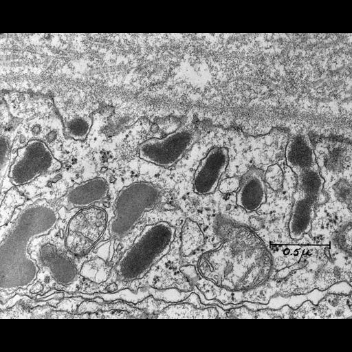
Stimulated Exocytosis. This micrograph shows the peripheral cytoplasm of a mammotroph in the pituitary of a lactating rat. Several secretion granules are lined up facing the perivascular spaces and are undergoing regulated exocytosis (The companion image (SFC-2a) that is grouped with this one (SFC-2) contains arrows labeling the exocytosis events.) The membrane of these secretion granules is in direct continuity with the cell membrane. The collagen in the extracellular matrix structure is evident. Bar=0.5 microns. This micrograph is one of the earliest to show the fusion of the membrane of secretory granules with the plasma membrane during the process of regulated secretion in endocrine cells. The pituitary of a lactating female rat was perfused with chilled 1.5% glutaraldehyde in 0.067 M cacodaylate buffer, pH 7.4, which contained 1% sucrose. The pituitary was removed from the rat and placed in fresh fixative for an additional 2 hours. Postfixation was with 1% osmium tetroxide in acetate-veronal buffer, pH 7.4. The sample was dehydrated and embedded in Epon. Sections were stained with uranyl acetate followed by lead citrate. Lysosome function in regulating secretion: disposal of secretory granules in cells of the anterior pituitary gland. In: Dingel JT, Fell HB, eds. Lysosomes in Biology and Pathology. Amsterdam: North Holland Publishing Co.; 1969:462-482. This image was scanned as 1200 dpi tiff (3986 x 3243 pixels) from the original lantern slide (3.25 x 4 inches).
| Spatial Axis | Image Size | Pixel Size |
|---|---|---|
| X | 4800px | 0.61nm |
| Y | 3905px | 0.61nm |