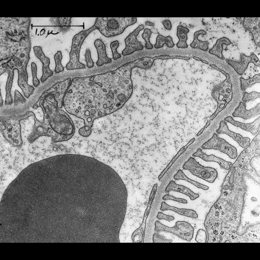
Peripheral area of normal rat kidney. The capillary wall is composed of three distinct layers: the endothelium with its fenestra, the basement membrane (BM) which is a continuous layer 0.1 to 0.15 microns in thickness and the foot processes of the renal epithelium. In a number of places (arrows), a slit membrane can be seen bridging the narrow gap between foot processes. The vascular lumen contains a red blood cell and ferritin. Image presented as a layered tiff with the first image the unlabeled image and the second image the labeled version. Bar= 1.0 microns. Demonstration of the structure of the filtration barrier of the normal kidney. Kidney was fixed by injection of 1% osmium tetroxide buffered in acetate-veronal, pH 7.5, into the renal parenchyma, the tissue was cut into small pieces and fixation was continued for 2 hours. Tissue blocks were embedded in Epon, sectioned and stained with lead hydroxide. Glomerular permeability investigated by electron microscopy. In: Siperstein MD, Colwell Sr. AR, Meyer K, eds. Small Blood Vessel Involvement in Diabetes Mellitus; Proceedings. Washington DC: American Institute of Biological Sciences; 1964:31-38.Pavenstadt H, Kriz W, Kretzler M. Cell biology of the glomerular podocyte. Physiol Rev [serial online]. 2003;83:253-307. EM Annotator: Marilyn G Farquhar (University of California, San Diego, CA). EM Annotator: Kathryn E Howell (University of Colorado School of Medicine, Aurora, CO). Original resource provided by Marilyn G Farquhar. Original resource: lantern slide (3.25 x 4 inches).
| Spatial Axis | Image Size | Pixel Size |
|---|---|---|
| X | 4800px | 1.03nm |
| Y | 4170px | 1.03nm |