Licensing

Pd
Public Domain: This image is in the public domain and thus free of any copyright restrictions. However, as is the norm in scientific publishing and as a matter of courtesy, any user should credit the content provider for any public or private use of this image whenever possible. Learn more
Comments: Powered by Pnyxe

Description

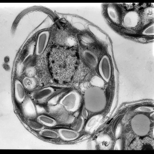
Appriximately longitudinal section of the unicellular alga Chlamydomonas reinhardtii showing one of the paired flagella, and the spherical nucleus surrounded by the single cup-shaped chloroplasts containing storage granules.

Technical Details

250 nm 'thick' epon section imaged with a high voltage JEM-1000 TEM at 1000KV.

Biological Sources

NCBI Organism Classification
Chlamydomonas reinhardtii
Cell Type
plant cell
Cell Line
strain in culture
Cellular Component
chloroplast
nucleus
microtubule-based flagellum part
microtubule basal body

Biological Context

Biological Process
photosynthesis, light harvesting
organelle organization

Attribution

Names
Mary Morphew
J. Richard McIntosh

Grouping

This image is part of a group.

Imaging

Image Type
transmission electron microscopy (TEM)
film
Imaging Mode
FBbi:00000622
detection of electrons
Parameters Imaged
electron density
Source of Contrast
elastic scattering of electrons
Visualization Methods
uranyl salt
Processing History
unprocessed raw data

Sample Preparation

Methods
glutaraldehyde fixed tissue
tissue in plastic embedment
Relation To Intact Cell
microtome-sectioned tissue

Dimensions

Spatial Axis Image Size Pixel Size
X 4862px 1.5nm
Y 4696px 1.5nm
*CIL – Cell Image Library accession number. Please use this to reference an image.