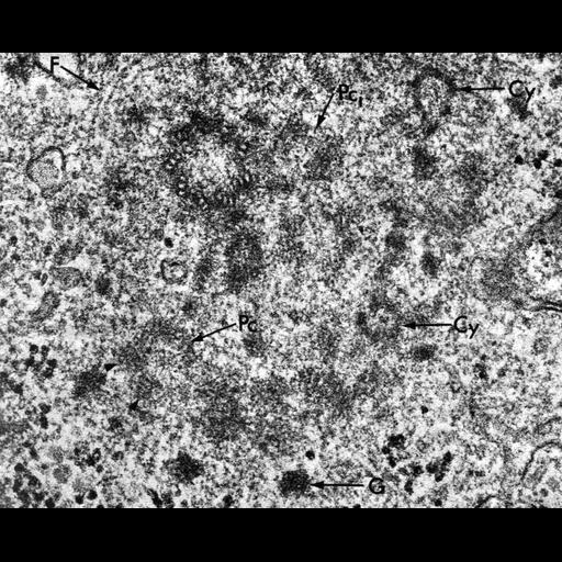
Cross-section of one of the centrioles of a diplosome and the surrounding region. The cells of a ciliated epithelium often produce many cilia, each of which grows from a 'basal body,' a structure that looks very much like a centriole. Basal bodies form in association with centrioles by a complex process that is readily visualized in the electron microscope, although the underlying biochemistry of basal body formation is not obvious from these images. One complete centriole is visible here as a ring of 9 triplet microtubules. Adjacent to it is a 'procentriole' (Pc1), which is an early stage in the formation of a new centriole. Pc indicates another procentriole, and cylinders (Cy) represent additional forming centrioles lying nearby. Dense granules (G) of unknown composition are common in this region of the cell. This micrograph, others from the same paper, and contemporary work by Ruth V Dippel described the remarkable diversity of structures that formed in association with existing centrioles as the many basal bodies needed to form multiple cilia formed in differentiating ciliated epithelia.
Trachea were dissected from chick embryos 15 to 19 days post fertilization, fixed in 3% glutaraldehyde, then 1% OsO4. Samples were then dehydrated in ethanol, embedded in epoxy plastic, sectioned, double-stained with uranyl acetate and lead citrate and imaged in an electron microscope (Siemens-Elmiskop 1). Image is at 87,000x and is Figure 8 in Kalnins and Porter. Micrograph was created in the mid-1960s while Vitauts I Kalnins was a member of the Laboratory of Keith Porter at Harvard. Original resource provided by Keith R Porter Archives (University of Maryland Baltimore County, Baltimore, MD). Scanned from published article at 1200 dpi and saved as a tif file.
| Spatial Axis | Image Size | Pixel Size |
|---|---|---|
| X | 4500px | 21.1667µm |
| Y | 3654px | 21.1667µm |