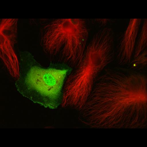
Sub-confluent, interphase Human Umbilical Vein Endothelial Cells (HUVECs) transfected with GFP-WT-MCAK (Mitotic Centromere Associated Kinesin) and grown in culture for 8 hours. Cells were fixed in PHEM buffer containing 4% paraformaldehyde, 0.05% gluteraldehyde, and 0.2% Triton X-100. Cells were then labeled with mouse anti-tubulin detected by goat anti-mouse Alexa 568 antibody. Cells were imaged on a spinning disk confocal microscope using a 60x 1.4 NA oil immersion objective lens on a Nikon Eclipse Ti equipped with Perfect Focus System and a Yokogawa CSU-X spinning disk confocal scan head equipped with a multi-bandpass dichromatic mirror (Semrock; Rochester, NY) and bandpass filters (Chroma; Rockingham, VT) in an electronic filterwheel for selection of GFP or Texas red emission. Excitation light provided by a custom-built laser combiner module (modification of LMM-3, Spectral Applied Research; Richmond Hill, Ontario, Canada) containing 500mW solid state lasers (488 nm: Coherent; 561 nm: MPB Communications; Montreal, Quebec) that were shuttered with electronic shutters and attenuated and/or directed to a fiber-coupled output port with an AOTF (Neos Technologies, Melbourne, FL) and directed to the confocal scan-head via a single-mode optical fiber (Oz Optics, Ottawa, Ontario, Canada). A Coolsnap HQ2 camera captured images with 200ms exposure @ 488nm and 400ms exposure @ 560nm.
| Spatial Axis | Image Size | Pixel Size |
|---|---|---|
| X | 1392px | 105µm |
| Y | 1040px | 105µm |
| Channel | Wavelength | |
|---|---|---|
| 1 | 488nm | |
| 2 | 561nm |