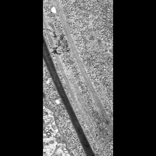
A high resolution, longitudinal section through one toxicyst and one bundle of microtubules (nematodesma) of the oral area of Didinium involved in the killing and ingesting of large prey. Didinium specializes in consuming Paramecium, an organism nearly their own size in less than one minute at standard room temperature. This slightly oblique longitudinal section allows the opportunity to visualize the longitudinal substructure of the hexagonally packed microtubules that support the cytopharynx and assistin the process. TEM taken on 5/20/69 by R. Allen with Philips 300 operating at 60kV. Neg. 6,370X. The raw film was scanned with a Nikon Coolscan 9000ED. This image is suitable for quantitative analysis. Standard glutaraldehyde fixation followed by osmium tetroxide, dehydrated in alcohol and embedded in an epoxy resin. Microtome sections prepared at approximately 75nm thickness. Additional information available at (http://www5.pbrc.hawaii.edu/allen/).
| Spatial Axis | Image Size | Pixel Size |
|---|---|---|
| X | 3504px | 0.83nm |
| Y | 7801px | 0.83nm |