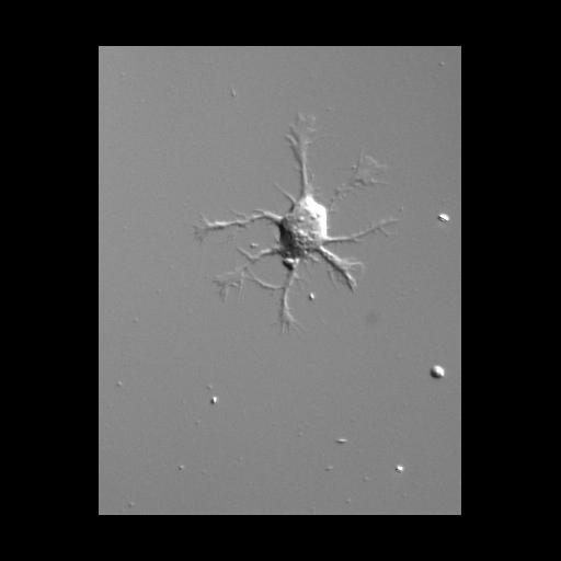
Time lapse recording showing the development of neuron polarity in a hippocampal neuron growing in vitro. This recording captures not only the formation of the axon (the longest process, oriented downward in the last frame), but also accelerated development and outgrowth of the dendritic arbor due to treatment with BMP-7, a growth factor that has been found to selectively promote dendritic development in these cells [as described in Withers et al., (2000) PMID: 10651865]. The coverslip containing neurons was mounted in a sealed temperature controlled chamber (Warner Instruments) in phenol-free Minimal Essential Medium supplemented with N2, pyruvate, and 30 ng/ml human recombinant BMP-7. Recording was made using an inverted Leica DMIRBE microscope, 40X PL Fluotar, 0.75 N.A. objective, Princeton Instruments MicroMax camera, and MetaMorph software. Images were acquired every 5 minutes, beginning 5 hours after plating, and continuing for over 19 hours.
| Spatial Axis | Image Size | Pixel Size |
|---|---|---|
| X | 331px | 0.169µm |
| Y | 428px | 0.169µm |
| Time | 300 seconds | 19 hr 20 min |
|---|