Alternate header for print version


Licensing
This image is copyright protected. Any public or private use of this image is subject to prevailing copyright laws. Please contact the content provider of this image for permission requests.
tweet  
*CIL – Cell Image Library accession number. Please use this to reference an image.

CIL:6568*  Cite 
Description

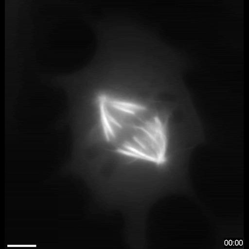
Mitotic spindle flattening. Compression elongates microtubule spindle. Widefield fluorescence images of Ptk2 cell expressing GFP tubulin flattened with a micromanipulator at 3:00-3:30 (min:s). Imaged with a 60X 1.4 NA objective on a Nikon TE300. Images captured with a Hamamatsu Orca ER. Filter set was HQ FITC Chroma, Ex 480/40 and Em 535/550. Bar is 5 microns. Corresponds to Video 1 in Curr Biol. 2009 Jul 14;19(13):1086-95

Biological Sources
NCBI Organism Classification
Macropus sp.
Cell Type
epithelial cell
Cell Line
PTK2
Cellular Component
spindle microtubule
Biological Context
Biological Process
mitotic spindle organization
Attribution
Names
Sophie Dumont
Timothy J Mitchison
Published
Curr Biol. 2009 Jul 14;19(13):1086-95
Pubmed
19540117
Citation
Digital Object Identifier (DOI)
doi:10.7295/W9CIL6568
Archival Resource Key (ARK)
ark:/b7295/w9cil6568
Imaging
Image Type
recorded image
Image Mode
fluorescence microscopy
time lapse microscopy
Parameters Imaged
fluorescence emission
Source of Contrast
distribution of a specific protein
Visualization Methods
EGFP
Processing History
unprocessed raw data
Data Qualifiers
raw, unprocessed data
suitable for spatial measurements
Sample Preparation
Methods
unfixed tissue
Relation To Intact Cell
dispersed cells in vitro
Dimensions
Spatial Axis Image Size Pixel Size
X 571px 0.2µm
Y 595px 0.2µm
Time 30 seconds