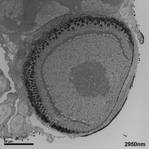
This study of the zebrafish eye is a series of four images taken at increasing magnifications. This image, the first image is the SEM block face of the zebrafish eye. This is followed by a second image that is a higher magnification TEM image of the plexiform layer and the outer nuclear layer of the zebrafish retina. The third image is a 15,000X TEM image of the bipolar and horizontal cells interdigitating in the OPL layer of the zebrafish retina. The fourth image in the series is a higher magnification (60,000X) of the dense synaptic bar and synaptic vesicles. This image of the larval zebrafish eye 10dpf was collected on a Gatan 3View® serial block face scanning electron microscope (SBFSEM). The 3View® is attached to a FEI Quanta 600 FEG SEM. Block face imaging is a way to look at epon embedded specimens using backscattered electrons in the SEM. A diamond knife in the vacuum chamber of the SEM cuts hundreds of ultrathin serial sections, thus gathering a perfected registered stack of images. Reference for SBFSEM: Denk, W. and H. Horstmann. Serial block face scanning electron microscopy to reconstruct three- dimensional tissue nanostructure. PloS Biology vol 2, 11; e329 doi:10.1371/journal.pbio.0020329 Microwave processing, osmium tetroxoxide/osmid procedure for enhancing membranes, and copper lead en bloc staining. Procedure for specimen preparation available at http://www.stanford.edu/~redhair/JoAnn_Buchanan.
| Spatial Axis | Image Size | Pixel Size |
|---|---|---|
| X | 750px | 0.333µm |
| Y | 750px | 0.333µm |