Alternate header for print version


Licensing
Attribution Only: This image is licensed under a Creative Commons Attribution License. View License Deed | View Legal Code
tweet  
*CIL – Cell Image Library accession number. Please use this to reference an image.

CIL:48102*  Cite 
Description

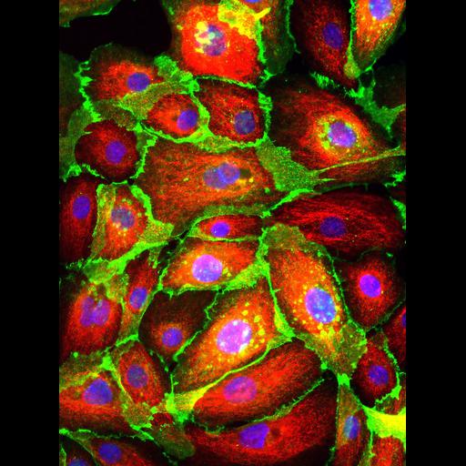
Cryopreserved human mammary epithelial cells were revived and stained for cytokeratin 18 (in red) to reveal the keratin-containing intermediate filaments found in the intracytoplasmic cytoskeleton of epithelial tissue, E-cadherin (in green) to visualize the calcium-dependent cell-cell adhesion glycoprotein, and DAPI (in blue) to label nuclei.

Technical Details

Cryopreserved human mammary epithelial cells were fixed and stained for cytokeratin 18, E-cadherin (from BD Transduction), and DAPI (from Life Technologies). Images were acquired with a 40X (Plan Fluor, NA 0.75) lens, SPOT RT Slider CCD camera and SPOT Basic image capture software. Image generated with the Image J color combine function.

Biological Sources
NCBI Organism Classification
Homo sapiens
Cell Type
mammary epithelial cell
Cellular Component
cytoskeleton
host cell membrane
nucleus
Biological Context
Biological Process
cell adhesion
Attribution
Names
Natalie Prigozhina
Citation
Digital Object Identifier (DOI)
doi:10.7295/W9CIL48102
Archival Resource Key (ARK)
ark:/b7295/w9cil48102
Imaging
Image Type
charge coupled device (CCD)
Image Mode
fluorescence microscopy
Parameters Imaged
fluorescence microscopy
Source of Contrast
distribution of epitope
Visualization Methods
4',6-diamidino-2-phenylindole (DAPI)
primary antibody plus labeled secondary antibody
Data Qualifiers
processed data
Dimensions
Spatial Axis Image Size Pixel Size
X 1600px ——
Y 1200px ——