Alternate header for print version


Licensing
Attribution Non-Commercial Share Alike:This image is licensed under a Creative Commons Attribution, Non-Commercial Share Alike License. View License Deed | View Legal Code
tweet  
*CIL – Cell Image Library accession number. Please use this to reference an image.

CIL:47812*  Cite 
Description

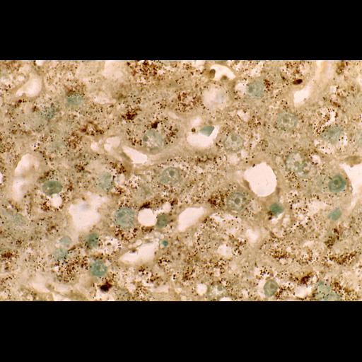
Bright-field image of human iiver parenchymal cells, where peroxisomes are stained dark brown with DAB by their catalase activity at pH 10.5, and postosmicated. Cell nuclei are counterstained with light green. Prefixation in buffered formal-calcium, cryostat section of 5 µm.

Technical Details

Normal image of human liver peroxisomes. Bright field light microscopy, immersion lens 63x.

Biological Sources
NCBI Organism Classification
Homo sapiens
Cell Type
parenchymal cell
Cellular Component
peroxisome
Biological Context
Molecular Function
catalase activity
Attribution
Names
Frank Roels
Citation
Digital Object Identifier (DOI)
doi:10.7295/W9CIL47812
Archival Resource Key (ARK)
ark:/b7295/w9cil47812
Imaging
Image Mode
bright-field microscopy
Source of Contrast
D