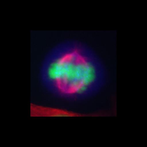
Shown is a fluorescence microscopy image of a HeLa cell at mitotic metaphase. The cell is stained to reveal DNA (blue), microtubules (red), and the C-terminal portion of the KIF2A protein (green) which is largely confined to the compact chromosomes. The image is part of a large project to characterize the locations of recombinantly tagged kinesin and myosin genes expressed from human or mouse-derived bacterial artificial chromosomes (BACs) as a resource for cell biology research. See also: Maliga et al. 2013 A genomic toolkit to investigate kinesin and myosin motor funtion in cells. Nature Cell Biol; published online Feb 2013;DCI:10.1038/ncb2689.
HeLa cells carrying the appropriate EGFP-labeled transgene were fixed in cold methanol, rehydrated, and treated with mouse antibody to alpha-tubulin and goat antibody to GFP followed by donkey-anti-mouse Alexa 594, donkey-anti-goat Alexa 488 and Hoechst 33342. Preparations were examined using a Zeiss Axioplan 2 widefield microscope with a 63x NA 1.4 objective lens. Images were recorded using a CCD camera (CS472-95, Hamamatsu). For further details see: Zaliga M et al. 2013 A genomic toolkit to investigate kinesin and myosin motor funtion in cells. Nat. Cell Biol. published online Feb 2013;DCI:10.1038/ncb2689.
| Spatial Axis | Image Size | Pixel Size |
|---|---|---|
| X | 300px | 0.106µm |
| Y | 300px | 0.106µm |