Alternate header for print version


Licensing
Attribution Non-Commercial; No Derivatives:This image is licensed under a Creative Commons Attribution, Non-Commercial, No Derivatives License. View License Deed | View Legal Code
tweet  
*CIL – Cell Image Library accession number. Please use this to reference an image.

CIL:42516*  Cite 
Description

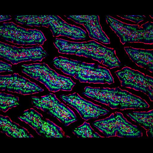
Fluorescence micrograph of the gut of transgenic GFP rat showing actin (red) and nuclei (blue). Image taken with a 20X objective. Honorable mention, 2004 Olympus BioScapes Digital Imaging Competition®.

Technical Details

Biological Sources
NCBI Organism Classification
Rattus
Cellular Component
actin cytoskeleton
nucleus
Biological Context
Mouse Gross Anatomy
TS25,gut
Attribution
Names
Mark Ross
2004 Olympus BioScapes Digital Imaging Competition®
Link
2004 Olympus BioScapes Digital Imaging Compet...
OTHER
University of Pittsburgh School of Medicine, Center for Biologic Imaging Pittsburgh, PA, USA
Citation
Digital Object Identifier (DOI)
doi:10.7295/W9CIL42516
Archival Resource Key (ARK)
ark:/b7295/w9cil42516
Imaging
Image Type
recorded image
Image Mode
fluorescence microscopy
Parameters Imaged
fluorescence emission
Source of Contrast
distribution of a specific protein
distribution of epitope
Visualization Methods
EGFP
Sample Preparation
Relation To Intact Cell
sectioned tissue
Dimensions
Spatial Axis Image Size Pixel Size
X 1280px ——
Y 1024px ——