Alternate header for print version


Licensing
Public Domain: This image is in the public domain and thus free of any copyright restrictions. However, as is the norm in scientific publishing and as a matter of courtesy, any user should credit the content provider for any public or private use of this image whenever possible. Learn more
tweet  
*CIL – Cell Image Library accession number. Please use this to reference an image.

CIL:42451*  Cite 
Description

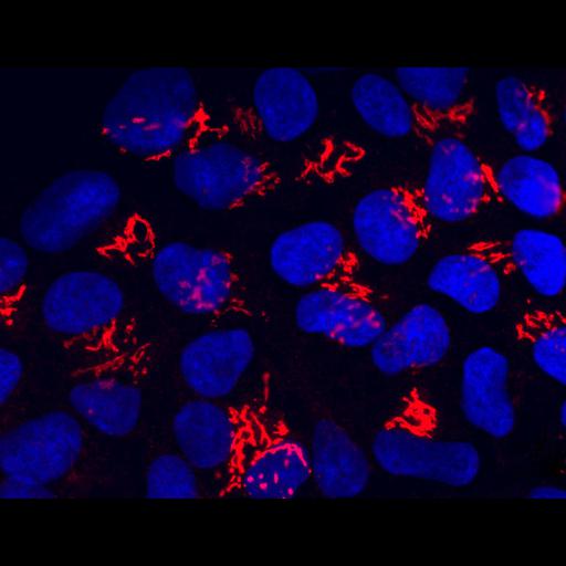
Immunofluorescence image of Human Embryonic Stem cells stained with an antibody to the active form of Bax (red) and Hoechst (blue). The distribution of Bax, an apoptosis mediator indicates that it resides in the Golgi apparatus under these conditions.

Technical Details

Cells were fixed with formaldehyde, permeabilized using Triton X-100, and stained with anti-Bax antibody 6A7 which is specific to the active form (red) and Hoechst dye to stain nuclei (red). Images were recorded on a Hamamatsu ORCA-ER digital camera mounted on a Leica DMIRE2 inverted microscope. See also: Dumitru R, Gama V, Fagan BM, Bower JJ, Swahari V, Pevny LH, Deshmukh M. 2012. Human embryonic stem cells have constitutively active Bax at the Golgi and are primed to undergo rapid apoptosis. Mol Cell. 46(5):573-83.

Biological Sources
NCBI Organism Classification
Homo sapiens
Cell Type
embryonic stem cell
Cellular Component
nucleus
Golgi apparatus
Biological Context
Biological Process
apoptosis
Attribution
Names
M. Deshmukh
R. Dimitru
V. Gama
B.M. Fagan
J.J. Bower
V. Swahari
L.H. Pevney
Link
NIGMS Biomedical Beat
Citation
Digital Object Identifier (DOI)
doi:10.7295/W9CIL42451
Archival Resource Key (ARK)
ark:/b7295/w9cil42451
Imaging
Image Type
charge coupled device (CCD)
Image Mode
fluorescence microscopy
Parameters Imaged
fluorescence emission
Source of Contrast
distribution of epitope
differences in adsorption or binding of stain
Visualization Methods
Hoechst 33258
Alexa Fluor 488
Data Qualifiers
processed data
Sample Preparation
Methods
formaldehyde fixed tissue
Relation To Intact Cell
whole mounted tissue
Dimensions
Spatial Axis Image Size Pixel Size
X 1344px ——
Y 1024px ——