Alternate header for print version


Licensing
Attribution Non-Commercial; No Derivatives:This image is licensed under a Creative Commons Attribution, Non-Commercial, No Derivatives License. View License Deed | View Legal Code
tweet  
*CIL – Cell Image Library accession number. Please use this to reference an image.

CIL:42003*  Cite 
Description

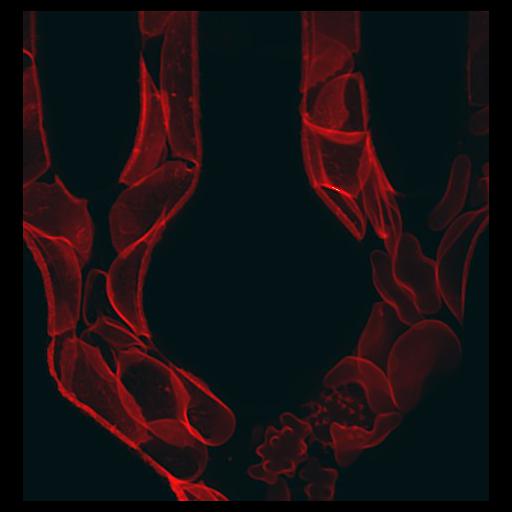
Confocal image of sickle red blood cells. To learn more about how sickle cells can clog blood vessels, their flow is observed in a microfabricated device designed to mimic a capillary bed in vivo. The misshapen sicklecells become lodged and occlude the microchannels of the device. Ross Rounsevell and Wilbur Lam, Department of Bioengineering, University of California, Berkeley, USA. Honorable Mention, 2009 Olympus BioScapes Digital Imaging Competition®.

Technical Details

Biological Sources
Cell Type
blood cell
Cellular Component
cell surface
Biological Context
Biological Process
blood circulation
Human Disease
sickle cell anemia
Attribution
Names
Ross Rounsevell
Wilbur Lam
2009 Olympus BioScapes Digital Imaging Competition®
Link
2009 Olympus BioScapes Digital Imaging Compe...
OTHER
Department of Bioengineering, University of California, Berkeley, USA
Citation
Digital Object Identifier (DOI)
doi:10.7295/W9CIL42003
Archival Resource Key (ARK)
ark:/b7295/w9cil42003
Imaging
Image Type
recorded image
Image Mode
confocal microscopy
Parameters Imaged
fluorescence emission
Sample Preparation
Methods
living tissue
Relation To Intact Cell
isolated cells
Dimensions
Spatial Axis Image Size Pixel Size
X 466px ——
Y 490px ——