Alternate header for print version


Licensing
Attribution Non-Commercial; No Derivatives:This image is licensed under a Creative Commons Attribution, Non-Commercial, No Derivatives License. View License Deed | View Legal Code
tweet  
*CIL – Cell Image Library accession number. Please use this to reference an image.

CIL:41825*  Cite 
Description

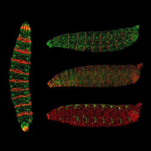
Confocal image stacks of Drosophila (fruit fly) larvae with labeled dendrite arborization sensory neurons and epidermal cells. In the vertical larva, green is dendrites of sensory neurons on the body wall and red is the posterior half of epidermal cells of every segment. In the horizontal larvae, in the top larva green is dendrites of sensory neurons on the body wall and red is attachment sites of body wall muscles. In the middle larva green is cell borders of epidermal cells and red is dendrites of sensory neurons on the body wall. In the bottom larva green and red are dendrites of two types of sensory neurons on the body wall. Honorable Mention, 2010 Olympus BioScapes Digital Imaging Competition®.

Technical Details

Biological Sources
NCBI Organism Classification
Drosophila melanogaster
Cell Type
epidermal cell
sensory neuron
Cellular Component
neuron projection
Biological Context
Biological Process
sensory neuron organization
Attribution
Names
Chun Han
2010 Olympus BioScapes Digital Imaging Competition®
Link
2010 Olympus BioScapes Digital Imaging Compe...
OTHER
University of California, San Francisco, CA, USA
Citation
Digital Object Identifier (DOI)
doi:10.7295/W9CIL41825
Archival Resource Key (ARK)
ark:/b7295/w9cil41825
Grouping This image is part of a group.
Imaging
Image Type
recorded image
Image Mode
confocal microscopy
Parameters Imaged
fluorescence emission
Processing History
projection of confocal stacks
Sample Preparation
Relation To Intact Cell
whole mounted tissue
Dimensions
Spatial Axis Image Size Pixel Size
X 2750px ——
Y 2341px ——