Alternate header for print version


Licensing
Attribution Non-Commercial; No Derivatives:This image is licensed under a Creative Commons Attribution, Non-Commercial, No Derivatives License. View License Deed | View Legal Code
tweet  
*CIL – Cell Image Library accession number. Please use this to reference an image.

CIL:41633*  Cite 
Description

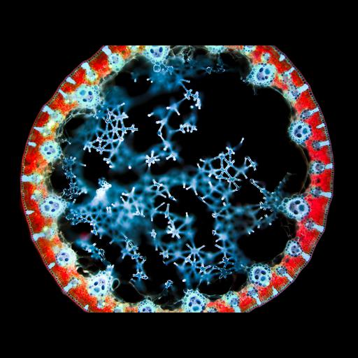
Fluorescence micrograph showing the cross-section of bulrush (Juncus sp.) leaf, autofluorescing red (chlorophyll on external side of leaf) and blue (vascular bundles). The diameter of the stalk is approximately 3mm.Honorable Mention, 2011 Olympus BioScapes Digital Imaging Competition®.

Technical Details

Biological Sources
NCBI Organism Classification
Juncus
Cellular Component
chlorophyll
Biological Context
Biological Process
leaf morphogenesis
Plant Growth
vascular bundle
leaf
Attribution
Names
Jan Martinek
2011 Olympus BioScapes Digital Imaging Competition®
Link
2011 Olympus BioScapes Digital Imaging Compe...
OTHER
Ostrov, Czech Republic
Citation
Digital Object Identifier (DOI)
doi:10.7295/W9CIL41633
Archival Resource Key (ARK)
ark:/b7295/w9cil41633
Imaging
Image Type
recorded image
Image Mode
fluorescence microscopy
Parameters Imaged
fluorescence emission
Source of Contrast
autofluorescence
Sample Preparation
Relation To Intact Cell
sectioned tissue
Dimensions
Spatial Axis Image Size Pixel Size
X 4140px ——
Y 3096px ——