Alternate header for print version


Licensing
Public Domain: This image is in the public domain and thus free of any copyright restrictions. However, as is the norm in scientific publishing and as a matter of courtesy, any user should credit the content provider for any public or private use of this image whenever possible. Learn more
tweet  
*CIL – Cell Image Library accession number. Please use this to reference an image.

CIL:40673*  Cite 
Description

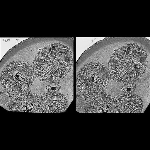
Stereo pair of an embedment-free section of a detergent-extracted 8-cell embryo reveals the cytoskeletal interior in greater detail than is afforded in resin-embedded sections. The specimen is viewed at an angle by selecting a stereo pair of -12 and 6 degrees (left to right), consequently the bottom left corner appears lower in the stereomicrograph. At later stages of development the specialized intermediate filament network in mammalian eggs and embryos (i.e., the cytoskeletal whorls) become parallel bundles as they are preparing to disassemble into individual intermediate filaments. Scale bar is 1.0 µm.

Technical Details

Sections were imaged with a Phillips CM300 transmission electron microscope. See Capco DG, McGaughey RW. Dev Biol. 1986 Jun;115(2):446-58 for details on specimen processing.

Biological Sources
NCBI Organism Classification
Mesocricetus auratus
Cell Type
early embryonic cell
Cellular Component
intermediate filament cytoskeleton
Biological Context
Biological Process
intermediate filament cytoskeleton organization
Attribution
Names
James J Faust
David G. Capco
OTHER
Arizona State University, Tempe, AZ
Citation
Digital Object Identifier (DOI)
doi:10.7295/W9CIL40673
Archival Resource Key (ARK)
ark:/b7295/w9cil40673
Grouping This image is part of a group.
Imaging
Image Type
recorded image
Image Mode
transmission electron microscopy (TEM)
Parameters Imaged
detection of electrons
Source of Contrast
differences in adsorption or binding of stain
Processing History
stereo pair
Sample Preparation
Methods
detergent permeabilized
chemically fixed tissue
osmium tetroxide fixed tissue
embedment medium removal
Relation To Intact Cell
sectioned tissue
Dimensions
Spatial Axis Image Size Pixel Size
X 814px ——
Y 414px ——