Licensing

Copyright
This image is copyright protected. Any public or private use of this image is subject to prevailing copyright laws. Please contact the content provider of this image for permission requests.
Comments: Powered by Pnyxe

Description

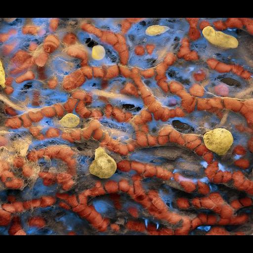
Colorized scanning electron micrograph showing the erythrocytes (red blood cells) within the capillary network of an alveola. The alveolar-capillary barrier (or membrane, or blood-air barrier) exists in the gas exchanging region of the lungs. It exists to prevent air bubbles from forming in the blood, and from blood entering the alveoli.

Technical Details

Image collected on a FEI Quanta Family with the following parameters: Magnification: 1800:1 x, Vacuum: High Vac, Voltage: 5 kv, Detector: SE+BSE, and Working Distance: 9 mm.

Biological Sources

Cell Type
blood cell
erythrocyte
Cellular Component
cell surface

Biological Context

Biological Process
gas transport
Human Development Anatomy
capillary
alveola

Attribution

Name
Oliver Meckes
Link
FEI Life Sciences Images

Imaging

Image Type
recorded image
Imaging Mode
scanning electron microscopy (SEM)
Parameters Imaged
elastic scattering of electrons
Visualization Methods
visualization of contiguous regions
Processing History
colorized

Sample Preparation

Relation To Intact Cell
whole mounted tissue

Dimensions

Spatial Axis Image Size Pixel Size
X 670px ——
Y 576px ——
*CIL – Cell Image Library accession number. Please use this to reference an image.