Alternate header for print version


Licensing
This image is copyright protected. Any public or private use of this image is subject to prevailing copyright laws. Please contact the content provider of this image for permission requests.
tweet  
*CIL – Cell Image Library accession number. Please use this to reference an image.

CIL:40592*  Cite 
Description

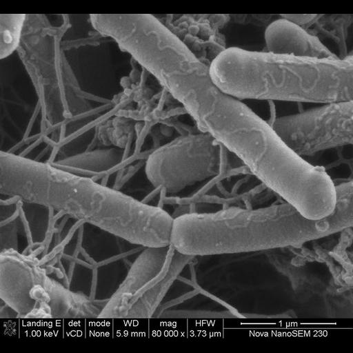
This scanning electron micrograph of salmonella bacteria, was made possible by using low kV to visualize the surface detail of the bacteria. The image was taken with the VcD (backscatter detector), using beam decelleration.

Technical Details

The image was collected on FEI instrument: Nova NanoSEM Family using the following parameters: Magnification: 80,000x, Horizontal Field Width: 3.73μm, Voltage: 1 kV, Detector: VcD, Spot: 2 nA, and Working Distance: 5.9 mm.

Biological Sources
NCBI Organism Classification
Salmonella
Cellular Component
cell surface
Biological Context
Biological Process
induction of bacterial agglutination
Attribution
Names
Miranda Waldron
Link
FEI Life Sciences Images
Citation
Digital Object Identifier (DOI)
doi:10.7295/W9CIL40592
Archival Resource Key (ARK)
ark:/b7295/w9cil40592
Imaging
Image Type
recorded image
Image Mode
back-scattered_electron imaging
Parameters Imaged
elastic scattering of electrons
Visualization Methods
visualization of contiguous regions
Processing History
unprocessed raw data
Sample Preparation
Relation To Intact Cell
whole mounted tissue
Dimensions
Spatial Axis Image Size Pixel Size
X 670px ——
Y 617px ——